Kamagra repose sur le sildénafil comme principe actif, avec un mode d’action identique à celui du Viagra. La forme galénique en gel oral permet une absorption plus rapide et une concentration plasmatique maximale plus précoce que les comprimés. Le mécanisme implique l’inhibition compétitive de la PDE5, entraînant une relaxation musculaire lisse locale et une vasodilatation ciblée. La demi-vie courte, environ 4 heures, limite la durée d’action. L’élimination se fait après métabolisme hépatique, impliquant majoritairement le CYP3A4. L’incidence d’effets indésirables comprend céphalées, rougeurs et congestion nasale, de façon transitoire. Dans les comparatifs pharmacologiques, acheter kamagra sans ordonnance est associé aux présentations galéniques alternatives disponibles.
03.pdf
JOP. J. Pancreas (Online) 2001; 2(4):140-149.Effect of Treatment with Different Doses of 17-β-Estradiol on Insulin Receptor Substrate-1. Celestino González, Ana Alonso, Natalia A Grueso, Fernando Díaz, Manuel M Esteban, Serafina Fernández, Angeles M Patterson
Department of Functional Biology, Physiology Area, University of Oviedo, Oviedo, Spain. ABSTRACT
levels in insulin dependent tissues, but in adifferent manner in each tissue. These novel
Context Ovarian hormones modulate insulin
sensitivity, but their exact role remains unclear.
knowledge about the possible risk for insulin
Objective We tried to determine whether
resistance in women taking oral contraceptives
different doses of 17-β-estradiol cause changes
or receiving hormone replacement therapy at
in the regulation of insulin receptor substrate
(IRS-1) levels, and if so, the possible implications in insulin sensitivity. Design Ovariectomized rats were treated with INTRODUCTION
different doses of 17-β-estradiol at 6, 11 and 16days.
Various clinical observations and experimental
Main outcome measures Immunoprecipitation
data suggest that estrogen and progesterone can
and Western blotting for IRS-1 were performed
modulate insulin sensitivity in females [1, 2]. In
Results We found that estradiol treatment has
pregnancy, where estrogen and progesterone
an influence on the amount of IRS-1 but that it
concentrations are markedly elevated, have a
acts in different ways depending on the tissue
substantial effect on carbohydrate metabolism
studied, on the length of treatment, and on the
as well as on the alteration of insulin sensitivity
[3]. Moreover, in women taking the combined
Conclusions Our results suggest that low
oral contraceptive pill, artificially increased
concentrations of 17-β-estradiol could be
levels of the sex steroid hormones estrogen and
responsible for the upregulation of insulin
progesterone were found to affect glucose
tolerance and insulin sensitivity [4, 5].
sensitivity in muscle and adipose tissue.
However, it remains unclear whether estrogens
alone or progestins alone can cause insulin
downregulated with high concentrations of 17-
resistance, or whether it is only a combination
β-estradiol, thus these high hormone plasma
levels could favour insulin resistance in
Insulin action begins upon its binding to its cell
peripheral tissues. The role of 17-β-estradiol
surface receptor [6, 7]. The discovery of
seems to modulate insulin receptor substrate 1
tyrosine kinase activity in the insulin receptor
JOP. Journal of the Pancreas – http://www.joplink.net – Vol.2, No.4 – July 2001
JOP. J. Pancreas (Online) 2001; 2(4):140-149.
implies that the mechanism of insulin action
of phosphorylation may also play a role in its
reduced response to insulin in the insulin-
transmitting the insulin signal. Insulin receptor
On the other hand, the possible contribution of
substrate 1 (IRS-1) is considered the major
estradiol to the degradation pathway of IRS-1 is
substrate for the insulin receptor, and contains
not sufficiently known, but the identification of
motifs that, after tyrosine phosphorylation, are
well-defined intermediates suggests that there
binding sites for proteins containing Src
may be a specific degradation pathway. Since
homology (SH2) domains [9]. The association
the role of IRS-1 is to act as a docking protein
of some proteins (phosphatidylinositol 3´-
which assembles a signaling complex, IRS-1
kinase: PI3-K, growth factor receptor-bound
protein 2: Grb2, and tyrosine phosphatase
SHP2) with IRS-1 permits their activation and
the transmission of the insulin signal [10].
demonstrate whether different doses of 17-β-
Thus, IRS proteins amplify the insulin receptor
estradiol could influence the regulation of the
amount of IRS-1 and the possible implication
constraints encountered by receptors that
of this hormone in insulin sensitivity during
directly recruit proteins containing SH2 site
gestation. Therefore, this study was designed to
domains to their autophosphorylation sites.
test whether the amount of IRS-1 in the liver,
Molloy et al. [11] and Lee et al. [12] showed
skeletal muscle and adipose tissue could be
that estrogen induces the expression of the
modified by the concentration of 17-β-estradiol
downstream signaling molecules, IRS-1 and
IRS-2. Estrogen induction of IRS-1 expression
was associated with increased tyrosinephosphorylation of IRS-1 and correlated with
MATERIAL AND METHODS
protein kinase (MAPK) activation. While the
increase in IRS-1 expression generally mirroredthe increase in tyrosine phosphorylation, the
Twelve-week-old virgin female Wistar rats
authors could not rule out the possibility that
(from the Biotery of the University of Oviedo)
the increase in tyrosine phosphorylation of IRS-
1 results from a change in stoichiometry or the
conditions of temperature (23±3 ºC) and
sites of phosphorylation. Furthermore, there
humidity (65±1%), with a regular lighting
schedule of a 12 h light/dark cycle (08:00 am -
08:00 pm) were used. The animals were fed a
decreased phosphorylation of IRS-1 may be
standard diet (Panlab A04, Barcelona, Spain)
mediated by the antiestrogen-induction of a
and had free access to water. All experimental
specific tyrosine phosphatase activity [13, 14].
manipulations were performed between 09:30
phosphorylation of IRS-1 on serine/threonineresidue has an inhibitory effect on insulin
Experimental Design
signaling [15, 16, 17]. Following the increase inIRS-1 serine/threonine phosphorylation, the
Three days before initiating the hormonal
ability of insulin to phosphorylate IRS-1 on
treatment (day -7), the rats were ovariectomized
tyrosine residues is decreased. It can be
through a midline incision using light ether
concluded, therefore, that a modulated pattern
anaesthesia. The ovariectomized rats were
JOP. Journal of the Pancreas – http://www.joplink.net – Vol.2, No.4 – July 2001
JOP. J. Pancreas (Online) 2001; 2(4):140-149.
randomly separated into four groups: control
to the temporal diagram reported in Table 1.
(V), estradiol (E), estradiol x 10 (EX10) and
Animals were sacrificed randomly on the 6th,
11th and 16th days (6 animals/subgroup). These
individually throughout the experiment.
days were selected as changes were found in
After surgery, the ovariectomized rats were
allowed 3 days to recover from the stress of
surgery and decrease their hormonal levels.
On the day of sacrifice and after 12 h of fasting
subcutaneously every twelve hours (09.00 am
anesthetized with 3.3 mL/kg body weight of
and 09:00 pm) for 20 days with 0.1 ml of a 17-
intraperitoneal ekytesin (0.96 g/mL sodium
β-estradiol (SIGMA Chemical Co., San Louis,
pentobarbital, 4.02 g/mL chloral hydrate, 2.12
USA) suspension in olive oil/ethanol (3:2 v/v).
g/mL magnesium sulphate, 40% propilenglicol,
The control group (V) injected with the vehicle
(olive oil/ethanol 3:2 v/v) was followed in
parallel. In the E group, different doses of 17-β-
reflexes, a blood sample (1 mL) was collected
estradiol were injected in order to simulate the
from the jugular vein in heparinized tubes,
centrifuged at 3000 rpm during 20 min at 4 ºC
pregnant rats [19, 20]. In the EX10 group, the
doses of 17-β-estradiol injected were ten times
stored frozen at –20 ºC until assayed. Plasma
E and in the EX100 group, the doses of 17-β-
17-β-estradiol was measured by RIA using
estradiol injected were one hundred times E.
The hormonal treatment was applied according
Biomedicals Inc., Costa Mesa, USA). The assaysensitivity was 10 pg/mL, and the intra-assay
Table 1. Temporal diagram of the 17- β-estradiol doses. Day 17-β-estradiol dose (E) Note
samples were measured on the same day. Each
Finally, samples of different tissues (liver;
skeletal muscle: flexor digitorum superficialis,extensor digitorum longus, soleus and extensordigitorum lateralis; retroperitoneal adipose
tissue) were collected and immediately frozen
in liquid nitrogen for future experiments and
the animals were killed by bleeding. Immunoprecipitation and Western Blotting
The samples of liver, skeletal muscle and
adipose tissue were washed with ice-cold sterile
Barcelona, Spain), 0.05% sodium deoxycholate,
JOP. Journal of the Pancreas – http://www.joplink.net – Vol.2, No.4 – July 2001
JOP. J. Pancreas (Online) 2001; 2(4):140-149.
speed for 30 sec. The extracts were centrifuged
(Western-Light, Chemiluminescent Detection
at 12,000 g at 4º C for 10 min in order to
System, TROPIX Inc., Bedford, USA) using a
remove insoluble material. After centrifugation,
1:2,000 dilution of polyclonal antibody against
the IRS-1 as the primary antibody, followed by
Bradford dye-binding method [21] using the
alkaline-phosphatase-conjugated anti-rabbit
Bio-Rad (Hercules, USA) reagents and BSA as
standard. The aqueous fraction containing 250
for detection. Finally, the membranes were
µg of protein for liver and muscle and 150 µg
rinsed several times with blocking buffer
without BSA and proteins were detected with
immunoprecipitation (IP) with 0.25 µg of
polyclonal antibody against the insulin receptor
Biotech, Barcelona, Spain) according to the
substrate 1 (IRS-1) (sc-559-G, Santa Cruz
Biotech, Inc., Santa Cruz, USA). The immune-
complexes were precipitated with protein G-
agarose beads (Roche Diag., Barcelona, Spain)
overnight at 4º C in a rocking platform and
Western blots were quantified using a digital
were washed several times in wash buffer (50
scanner (AX-110, Nikon, Surrey, UK) and NIH
Image 1.57 software (Scion Co., Maryland,
0.1% ortovanadate 1 M). After washing, thepellet was suspended in protein loading buffer
(250 mM Tris-HCl pH 6.8, 8% SDS, 8 mMEDTA, 35% glycerol, 2.5% β-mercapto-
The experiments were carried out in accordance
ethanol, Bromophenol Blue) and heated in a
with the rules of laboratory animal care.
boiling water bath for 5 min. For total extraction, similar size aliquots were
STATISTICS
subjected to SDS-PAGE (7% Tris-Acri-Bis) ina miniature slab gel apparatus (Bio-Rad,
Data are expressed as mean ± SEM. We used
Hercules, USA). The prestained molecular-
analysis of variance followed by a Tukey test in
mass standards used were myosin (218 kDa), β-
the statistical analysis of 17-β-estradiol plasma
galactosidase (126 kDa), bovine serum albumin
levels. Since the distribution of the IRS-1 levels
in different tissues was skewed, the Mann-
soybean trypsin inhibitor (33.9 kDa), lysozyme
(17.4 kDa) and aprotinin (7.6 kDa) (Bio-Rad,
used. A P value less than 0.05 was considered
Hercules, USA). Electrotransfer of proteins
significant. Statistical analysis was performed
(Hybond-ECL, Amersham Pharmacia Biotech,Barcelona, Spain) was performed for 60 min at
50 V (constant) in a miniature transferapparatus (Mini-Protean, Bio-Rad) as described
Plasma Levels of 17-β-Estradiol
by Towbin et al. [22]. Non-specific protein binding to the
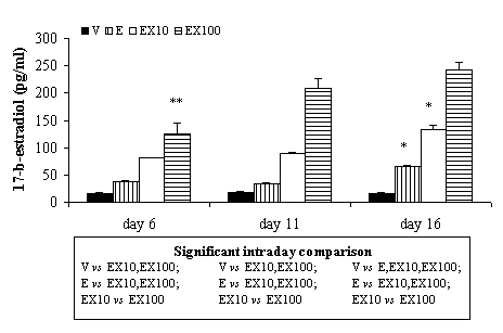
The plasma levels of 17-β-estradiol are shown
in Figure 1. The results obtained in the E group
preincubating the filter for two hours at room
were similar to normal pregnant rats at 5, 10
temperature in blocking buffer (TNT, 7% BSA)
and 15 days of pregnancy [19, 20]. In this
group, we found similar values at 6 and 11 days
JOP. Journal of the Pancreas – http://www.joplink.net – Vol.2, No.4 – July 2001
JOP. J. Pancreas (Online) 2001; 2(4):140-149.
In the liver, at day 6 of experimentation weobserved a significant increase in the IRS-1level in the E and the EX10 groups ascompared to the V and the EX100 groups. Thesame result was observed at day 11 but thedifference between the EX10 and the V groupsdid not reach the level of significance. At day16, we found a significant increase in the Egroup as compared to the other groups and asignificant decrease in the EX100 group ascompared to the EX10 group; no significantdifferences were found between the V and the
Figure 1. 17-β-estradiol plasma levels in ovariectomized
EX10 groups. The length of treatment does not
rats (V) and rats treated with different doses of 17-β-
significantly change the amount of IRS-1, but
estradiol (E, EX10, EX100). Data are expressed as mean
we observed a remarkable progressive increase
± SEM. Only significant differences are shown. Significant interday comparison: * day 16 vs. days 6 and
in the V group from day 6 to day 16 and a
remarkable decrease in the EX10 and theEX100 groups in the same period.
of treatment and a significant increase was
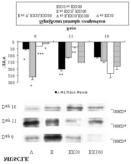
The level of IRS-1 in skeletal muscle was
observed at day 16 vs. days 6 and 11. The
significantly increased in the E group as
results obtained in the EX10 and the EX100
compared to the other groups at day 6.
groups were significantly higher than E.
However, at day 11 we found a significant
increase in the V group as compared to the
parallel with the E group. The 17-β-estradiol
other groups; we also showed that the EX100
plasma levels were dependent on the solution
group had significantly higher IRS-1 levels than
injected. Thus we observed that the plasma
the E and the EX10 groups and these levels
concentration of 17-β-estradiol increases: at
were significantly higher in the E group as
day 6 (238% in E vs. V, 509% in EX10 vs. V
compared to the EX10 group. Only significant
and 787% in EX100 vs. V), at day 11 (188% in
differences were found between V and EX10
E vs. V, 488% in EX10 vs. V and 1,145% in
groups at day 16. As far as the comparisons
EX100 vs. V) and at day 16 (401% in E vs. V,
between the different days were concerned, we
803% in EX10 vs. V and 1,463% in EX100 vs.
observed a significant increase of IRS-1 levels
in the V group from day 6 to day 11 and asignificant decrease from day 11 to day 16. In
Protein Content of IRS-1
the R group, the IRS-1 levels were significantlydecreased at days 11 and 16 vs. day 6, while the
Figure 2 shows a representative experiment in
length of treatment significantly increased these
which the solubilized liver, skeletal muscle and
levels in the EX10 (day 16 vs. days 6 and 11)
and the EX100 (days 11 and 16 vs. day 6)
antibody. After SDS-PAGE and electrotransfer,
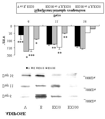
In adipose tissue, the level of IRS-1 was found
nitrocellulose membranes were incubated in the
to be significantly higher in the E and the EX10
groups as compared to the V group at day 6 of
immunoprecipitated materials, ECL detection
treatment. However, this tendency changes at
days 11 and 16 of treatment: the level of IRS-1
JOP. Journal of the Pancreas – http://www.joplink.net – Vol.2, No.4 – July 2001
JOP. J. Pancreas (Online) 2001; 2(4):140-149.Figure 2. IRS-1 levels in liver, skeletal muscle and adipose tissue in ovariectomized rats treated with different doses of 17-β-estradiol. The proteins were isolated, immunoprecipitated with anti-IRS-1 antibody and immunoblotted with anti-IRS-1 antibody. Scanning densitometry was performed for five independent experiments. Data are expressed as mean ± SEM. Only significant differences are shown. Significant interday comparison: * day 6 vs. days 11 and 16; ** day 11 vs. day 16; *** day 6 vs. day 16 A.S.U.: Arbitrary scanning units
between days 6 and 16, whereas in the EX10and the EX100 groups, the opposite occurred:this level decreased at day 16 vs. days 6 and 11in the EX10 group, as well as progressivelydecreasing during the treatment period in theEX100 group. However, in the E group wefound a significant decrease between days 6and 11 and a significant increase between days11 and 16.
compared to the other groups at days 11 and 16. We also observed that the tendency of the IRS-
DISCUSSION
1 levels throughout treatment was differentdepending on the timing and the 17-β-estradiol
We have recently showed that 17-β-estradiol
dose. In the V group, the level of IRS-1 rose
could be responsible for the increase in insulin
JOP. Journal of the Pancreas – http://www.joplink.net – Vol.2, No.4 – July 2001
JOP. J. Pancreas (Online) 2001; 2(4):140-149.
sensitivity during early pregnancy when the
These findings confirm a previous study which
plasma concentrations of 17-β-estradiol and
demonstrated that gestation could be divided
into two periods, namely, the period of early
pregnancy when the plasma concentrations of
gestation, which is characterized by an increase
both hormones are high, the role of 17-β-
in sensitivity to insulin action in the maternal
estradiol could be that of antagonizing the
tissues and the period of late gestation,
effect of progesterone by diminishing insulin
characterized by a decrease in this sensitivity
sensitivity. We therefore propose that some of
[19]. In the light of the present results our
hypothesis is that, during early pregnancy, the
metabolism during pregnancy can be focused
peripheral "insulin sensitivity" (larger amount
of IRS-1) together with high insulin plasma
The novel finding of this study is that 17-β-
levels (data not shown) could favour the storage
estradiol seems to be responsible for controlling
of energetic reserve in the adipose tissue,
insulin sensitivity in females throughout
gestation. In this sense, the present results
adaptative changes are reversed. The decrease
confirm those of a previous study [23], namely,
in insulin plasma levels (data not shown)
together with less insulin sensitivity in adipose
estradiol is similar to that found in early
tissue facilitate the lipolysis and the increase in
pregnancy, IRS-1 is upregulated and the insulin
sensitivity increases in the peripheral tissues
observed at the end of pregnancy [20].
(skeletal muscle and adipose). However, IRS-1
In relation to the EX10 and the EX100 groups,
we can note that the higher doses of 17-β-
concentration of this hormone is similar to that
estradiol (EX100 group) significantly decreased
found in late pregnancy, diminishing the insulin
the amount of IRS-1 with respect to the E group
sensitivity in peripheral tissues. We suggest, in
in the liver and adipose tissue. However, in
accordance with Lee et al. [12], that a low
skeletal muscle, this fact only occurs at day 6 of
concentration of 17-β-estradiol increases IRS-1
expression by a transcriptional mechanism
hormone action seems to amplify the decrease
because the IRS-1 promoter does have four
in IRS-1 levels in the EX100 group, also with
consensus half- estrogen response elements
the exception of skeletal muscle. Similar results
[24]. However, when the concentration of 17-β-
findings seem to demonstrate that liver and
complexes of estradiol receptor-estradiol to the
adipose tissue are the tissues most affected by
IRS-1 promoter could induce a decrease in IRS-
17-β-estradiol action with respect to the amount
1 expression, probably by displacement of other
of IRS-1, and that the effect of this hormone
transcription factors linked to the IRS-1
was dose and timing dependent in agreement
promoter. Among these factors, we could point
with other authors postulating that the effect of
out progesterone, lactogenic hormones, growth
hormone, etc, [1, 2, 3, 23, 25, 26].
metabolism depend on the type of estrogen,
In the present study, in the E group, the tissues
dose, length of treatment, etc. [4, 27, 28]. The
studied were found to perform differently. In
roles of liver and adipose tissue are very
this way, there are no significant changes in the
important during pregnancy, because the liver
treatment, but, in skeletal muscle and adipose
clearance [29] and adipose tissue is the most
tissue, a significant decrease was observed from
important energetic reserve. In this sense, our
day 6 to day 11 and from day 6 to day 16.
hypothesis, in the light of the present results, is
JOP. Journal of the Pancreas – http://www.joplink.net – Vol.2, No.4 – July 2001
JOP. J. Pancreas (Online) 2001; 2(4):140-149.
that during early pregnancy, the liver could bediscouraging insulin clearance, so the high
Received March 2nd, 2001 – Accepted June
insulin plasma levels (data not shown) together
with high sensitivity in adipose tissue couldfavour the storage of energetic reserve. Key words Insulin; Insulin Resistance; Rats
However, in late pregnancy, the role of the livercould be to increase insulin clearance, so the
Abbreviations Grb2: growth factor receptor-
decrease in insulin plasma levels (data not
bound protein 2; IP: immunoprecipitation; IRS-
shown) together with less insulin sensitivity in
adipose tissue facilitate the lypolisis and the
increase in triglycerides plasma levels which
can be observed at the end of pregnancy [20].
We consider that these novel findings are veryimportant for a better understanding of the role
Acknowledgements This study was supported
of estrogen during pregnancy. Moreover, these
by grants from the University of Oviedo (NP-
results also illustrate the great importance of
estrogen dosage and concentration as regards
Universidades e Investigación del Principado
glucose metabolism in hormonal replacement
de Asturias as part of the II Plan Regional de
therapy in women at menopause and in women
taking oral contraceptives. In summary, our present findings suggest that
Correspondence
low concentrations of 17-β-estradiol similar to
early pregnancy levels could be responsible for
the upregulation of IRS-1, increasing insulin
sensitivity in peripheral tissues (muscle and
downregulated with high concentrations of 17-
β-estradiol similar to late pregnancy, thus these
insulin resistance in the peripheral tissues.
Consequently, the role of 17-β-estradiol seems
to be to modulate the amount of IRS-1 ininsulin dependent tissues, but in a differentmanner in each tissue. In spite of this, the role
References
of this hormone could appear to be slightlyaltered in the presence of high plasma
concentrations of progesterone, lactogenic
H, Bakker A, Heine RJ. Induction of insulin
hormones and growth hormone, just as occurs
resistance by androgens and estrogens. J Clin
during normal pregnancy. Moreover, we think
that these novel findings are very important in
order to improve knowledge about the possible
risk for insulin resistance in women taking oral
Adipocyte insulin action during the normal
contraceptives or hormone replacement therapy
menstrual cycle. Hum Reprod 1996; 11:968-74.
JOP. Journal of the Pancreas – http://www.joplink.net – Vol.2, No.4 – July 2001
JOP. J. Pancreas (Online) 2001; 2(4):140-149.
3 Saad MJA, Maeda L, Brenelli SL, Carvalho
signaling in human breast cancer: estrogen
CRO, Paiva RS, Velloso LA. Defects in insulin
regulation of insulin receptor substrate-1
signal transduction in liver and muscle of
expression in vitro and in vivo. Mol Endocrinol
pregnant rats. Diabetologia 1997; 40:179-86.
13 Freiss G, Vignon F. Antiestrogens increase
4 Godsland IF, Walton C, Felton C, Proudler
protein tyrosine phosphatase activity in human
breast cancer cells. Mol Endocrinol 1994;
secretion, and metabolism in users of oral
contraceptives. J Clin Endocrinol Metab 1992;
14 Freiss G, Puech C, Vignon F. Extinction of
associated protein tyrosine phosphatase-1 in
metabolism in oral contraceptive users without
human breast cancer cells. Mol Endocrinol
Endocrinol Metabolism 1994; 79:1277-83.
15 Tanti JF, Grémeaux T, Van Obberghen E,
phosphorylation of insulin receptor substrate 1
pathological conditions. Annu Rev Med 1990;
modulates insulin receptor signaling. J Biol
16 De Fea K, Roth RA. Modulation of insulin
JM, Araki E, Wilden PA, et al. Structure of the
receptor substrate 1 tyrosine phosphorylation
insulin receptor substrate IRS-1 defines a
and function by mitogen-activated protein
unique signal transduction protein. Nature
kinase. J Biol Chem 1997; 272:31400-6.
8 White MF, Kahn CR. The insulin signaling
modulation of insulin receptor substrate-1
tyrosine phosphorylation requires serine 612. Biochemistry 1997; 36:12939-47. [97477343]
9 Sun XJ, Miralpeix M, Myers MG, GlasheenEM, Backer JM, Kahn CR, White MF.
Expression and function of IRS-1 in insulin
mechanism of insulin action in normal and
insulin-resistant states. Exp Clin Endocrinol
19 González C, Díaz F, Fernández S, Patterson
signaling system. Trends Biochem Sci 1994;
AM. Role of 17-β-estradiol and progesterone
11 Molloy CA, May FEB, Westley BR. Insulin
restriction (50%) in pregnant and non-pregnant
receptor substrate-1 expression is regulated by
rats. J Endocrinol Invest 1997; 20:397-403.
estrogen in the MCF-7 human breast cancer cell
line. J Biol Chem 2000; 275:12565-71.
20 González C, Díaz F, Fernández S, Patterson
AM. Pregnancy in rats and food restriction
12 Lee AV, Jackson JG, Gooch JL, Hilsenbeck
(50%): insulin response in relation to serum
lipids and lipoprotein levels. Nutr Research
D. Enhancement of insulin-like growth factor
JOP. Journal of the Pancreas – http://www.joplink.net – Vol.2, No.4 – July 2001
JOP. J. Pancreas (Online) 2001; 2(4):140-149.
transduction in rat tissues. Mol Cell Endocrinol
quantities utilizing the principle of protein dye
binding. Anal Biochem 1976; 72:248-54.
26 Kawai M, Kishi K. Adaptation of pancreatic
islet B-cells during the last third of pregnancy:
Electrophoretic transfer of proteins from
regulation of B-cell function and proliferation
polyacrylamide gels to nitrocellulose sheets.
Procedure and some applications. Proc Natl
Acad Sci USA 1979; 76: 4350-4. [80056736]
27 Kojima T, Lindheim SR, Duffy DM, Vijod
23 González C, Alonso A, Alvarez N, Díaz F,
MA, Stanczyk FZ, Lobo RA. Insulin sensitivity
Martínez M, Fernández S, Patterson AM. Role
of 17-β-estradiol and/or progesterone on insulin
ethinyl estradiol used in oral contraceptives.
sensitivity in the rat: implications during
Am J Obstet Gynecol 1993; 169:1540-4.
pregnancy. J Endocrinol 2000; 166:283-91.
28 Lindheim SR, Duffy DM, Kojima T, Vijod
24 Kato S, Tora l, Yamauchi J, Masushige S,
administration influences the effect of estrogen
estrogen response element of the ovalbumin
gene contains several half- palindromic 5´-
women. Fertil Steril 1994; 62:1176-80.
TGACC-3´ motifs acting synergistically. Cell
25 Thirone ACP, Carvalho CRO, Brenelli SL,
mechanism, products, and significance. Endocr
Velloso LA, Saad MJ. Effect of chronic growth
JOP. Journal of the Pancreas – http://www.joplink.net – Vol.2, No.4 – July 2001
5HTT : Et si le bonheur était affaire de longueur ? Et si, apprenant la mort de Juliette, Roméo n’avait pas mis fin à ses jours par romantisme mais à cause d’une défaillance chimique ? Shakespeare s’en retournerait sans doute dans sa tombe ! Pourtant, une équipe de chercheurs pense que le mélodrame serait en grande partie une affaire de chimie. En effet, une protéine - la 5HTT
Conducted at the Center for Experimental and Applied Skin Physiology and Clinical Research Center for Hair and Skin Physiology of the University Clinic Effectiveness Study Tests regarding the penetration of caffeine from a shampoo formula Following successful tests at the University of Jena on the hair organ culture model regarding the effi cacy of caff eine as a hair growt
 JOP. J. Pancreas (Online) 2001; 2(4):140-149.
In the liver, at day 6 of experimentation weobserved a significant increase in the IRS-1level in the E and the EX10 groups ascompared to the V and the EX100 groups. Thesame result was observed at day 11 but thedifference between the EX10 and the V groupsdid not reach the level of significance. At day16, we found a significant increase in the Egroup as compared to the other groups and asignificant decrease in the EX100 group ascompared to the EX10 group; no significantdifferences were found between the V and the
Figure 1. 17-β-estradiol plasma levels in ovariectomized
JOP. J. Pancreas (Online) 2001; 2(4):140-149.
In the liver, at day 6 of experimentation weobserved a significant increase in the IRS-1level in the E and the EX10 groups ascompared to the V and the EX100 groups. Thesame result was observed at day 11 but thedifference between the EX10 and the V groupsdid not reach the level of significance. At day16, we found a significant increase in the Egroup as compared to the other groups and asignificant decrease in the EX100 group ascompared to the EX10 group; no significantdifferences were found between the V and the
Figure 1. 17-β-estradiol plasma levels in ovariectomized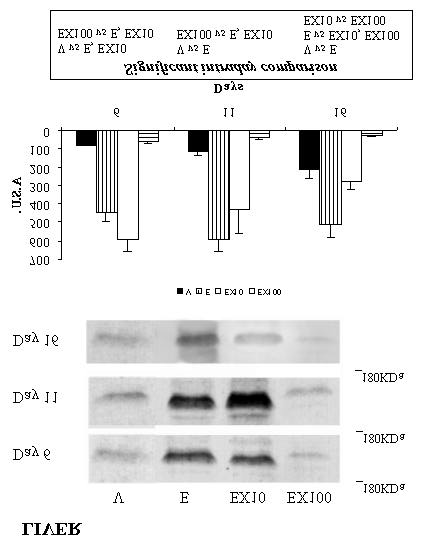
 JOP. J. Pancreas (Online) 2001; 2(4):140-149.
Figure 2. IRS-1 levels in liver, skeletal muscle and
JOP. J. Pancreas (Online) 2001; 2(4):140-149.
Figure 2. IRS-1 levels in liver, skeletal muscle and