Kamagra repose sur le sildénafil comme principe actif, avec un mode d’action identique à celui du Viagra. La forme galénique en gel oral permet une absorption plus rapide et une concentration plasmatique maximale plus précoce que les comprimés. Le mécanisme implique l’inhibition compétitive de la PDE5, entraînant une relaxation musculaire lisse locale et une vasodilatation ciblée. La demi-vie courte, environ 4 heures, limite la durée d’action. L’élimination se fait après métabolisme hépatique, impliquant majoritairement le CYP3A4. L’incidence d’effets indésirables comprend céphalées, rougeurs et congestion nasale, de façon transitoire. Dans les comparatifs pharmacologiques, acheter kamagra sans ordonnance est associé aux présentations galéniques alternatives disponibles.
Cadmium ion adsorption controls the growth of cds nanoparticles on layered montmorillonite and calumit surfaces
Journal of Colloid and Interface Science 213, 584 –591 (1999) Article ID jcis.1999.6174, available online at http://www.idealibrary.com on
Cadmium Ion Adsorption Controls the Growth of CdS Nanoparticles
on Layered Montmorillonite and Calumit Surfaces
I. De´ka`ny,* L. Turi,* G. Galba´cs,† and J. H. Fendler‡,1
*Department of Colloid Chemistry and Nanostructured Materials Research Group of the Hungarian Academy of Sciences;
†Department of Inorganic and Analytical Chemistry, Attila Jo´zsef University, Aradi V.t.1., H-6720 Szeged, Hungary; and
‡Center for Advanced Materials Processing, Clarkson University, Potsdam, New York 13699-5814
Received November 20, 1998; accepted February 17, 1999
trations in the nanoreactors have been assessed from adsorption
Adsorption isotherms have been determined for the intercala-
isotherms obtained in different ethanol– cyclohexane mixtures. tion of cadmium ions (Cd2؉) into layered hydrophobized montmo-
Information on the CdS particles formed has been deduced by
rillonite (HDP-M) and calumit (DBS-C) sheets dispersed in etha-
absorption spectrophotometric, small-angle X-ray diffraction
nol (1)– cyclohexane (2) mixtures. The amount of Cd2؉ adsorbed
(SAXS), and transmission electron microscopic (TEM) mea-
depended strongly on the composition of the binary liquid; at an ethanol mole fraction of 0.05 (x ؍ 0.05), 95% of the added Cd2؉ is located in the ethanolic nanoreactor at the HDP-M (or DBS-C) surface. CdS nanoparticles have been generated in situ in ethan- EXPERIMENTAL SECTION olic nanoreactors at the HDP-M and DBS-C surfaces. Absorption spectrophotometric measurements provided information on the number of CdS nanoparticles formed and on their absorption
The preparations, purifications, and characterizations of
edges, bandgaps, and mean diameters. Good correlations have
hexadecyl pyridium modified montmorrilonite, HDP-M, and
been obtained between the adsorption isotherms and the size (and
sodium dodecylbenzenesulfonate modified calumit, DBS-C,
the amount) of the CdS formed. X-ray diffractometry established that CdS nanoparticles stretched the HDP-M and DBS-C lamellas
have been described (5– 8). Ethanol and cyclohexane (p.a.,
unevenly upon intercalation.
Reanal, Hungary) were dried over 0.4 nm molecular sieves
1999 Academic Press Key Words: adsorption; nanoparticle growing; CdS; montmoril-
(Merck AG, Germany). Cadmium acetate (p.a., Reanal, Hun-
lonite; calumite; nanophase reactors.
gary) was used as received. Hydrogen sulfide was prepared, asneeded, from FeS and HCl in a Kipp apparatus (purified bywashing with aqueous NaOH and distilled water and dried over
INTRODUCTION
a passage through calcium carbonate).
The recognized importance of size quantization has
prompted the developments of viable routes for the preparation
2.1. Determination of cadmium ion adsorption.
of semiconductor nanoparticles in liquids and at solid inter-
acetate solubilities in adsorption on HDP-M and DBS-C were
faces (1– 4). We have successfully employed ultrathin polar
determined in a variety of different ethanol (1)– cyclohexane
liquid layers, selectively adsorbed on solid surfaces in contact
(2) mixtures. Dried (80°C) adsorbent (HDP-M or DBS-C),
with alcohol– cyclohexane binary mixtures, as nanoreactors for
0.05 g, was introduced into 50-mL ethanol (1)– cyclohexane
the preparation of semiconductor particles (1, 2). Specifically,
(2) mixture (containing the desired amount of x ) in a closed
we have generated CdS and ZnS nanoparticles in these nano-
glass container, dispersed by shaking and sonication (15 min in
reactors at montmorrilonite clay platelet (5, 6) and silica par-
a Realsonic RS-06 bath-type 100 sonicator), allowed to equil-
ibrate for 1 week, and centrifuged (20 min, 10,000 rpm).
Optimization of the process requires an understanding of the
Cadmium ions remaining in the supernatant (10 –20 mL) were
parameters which affect the formation of semiconductor par-
extracted into 30 mL, and their concentrations were determined
ticles. Attention is focused, therefore, in the present work on
by inductively coupled plasma atomic emission spectrometry
the influence of the ethanol to cyclohexane ratio and of the
(ICP-AES) to give the equilibrium concentration of cadmium
concentration of cadmium-ion precursors (cadmium acetate)
ion in the supernatant, C . This, in turn, permitted the assess-
on the size of the CdS nanoparticles, in situ formed on layered
ment of the amount of cadmium ion adsorbed, n s
montmorillonite and calumit surfaces. Cadmium ion concen-
To whom correspondence should be addressed.
ϭ V͑C Ϫ C ͒ma s,
0021-9797/99 $30.00Copyright 1999 by Academic PressAll rights of reproduction in any form reserved.
Cd2ϩ ADSORPTION CONTROLS CdS GROWTH IN CLAYS
The sample solutions were introduced to the nebulizer of the
ICP-AES spectrometer by a high-precision Gilson MinipulseIII peristaltic pump at a flow rate of 1.5 cm3/min. The argonplasma spectrometer was operated at 1 kW plasma forwardpower and 12 L/min plasma gas and 1 L/min sheath gas flowrate settings. Three-point linear calibration was performed,covering the range of concentration of interest, 1–100 ppm. The calibrating solutions were prepared by dilution of a cad-mium actetate stock solution (1 g/100 mL) with water. Ana-lytical line selection (226.502 nm Cd) and conventional two-point background correction were based on the inspection ofthe emission spectra of the samples. All measurements weretaken in triplicate, and concentrations were calculated withYobin-Yvon V 4.03 built-in data handling and software. 2.2. Preparation of CdS nanoparticles.
incorporated into layered HDP-M and DBS-C were preparedby the infusion of H S, in amounts equivalent to the Cd2ϩ ions
2.3. Determination of specific surface areas.
face areas of CdS–HDP-M and CdS DBS–C composites were
Solubility of Cd(Ac)2 in ethanol (1)– cyclohexane (2) mixtures at
determined by Micromeritics Gas Adsorption Analyzer (Gem-
25.0°C as a function of the ethanol mole fraction ( x 1).
ini Type 2375) at 77 K in liquid nitrogen. The adsorptionisotherms were analyzed with the BET equation.
where V is the volume of the total amount of liquid used, C is
the initial concentration, m is the mass of the adsorbent (in
determined on a Jobin-Yvon 24 sequential ICP-AES spectrom-
grams), and a s is the specific surface area of the adsorbent.
eter, equipped with a Babington-type nebulizer.
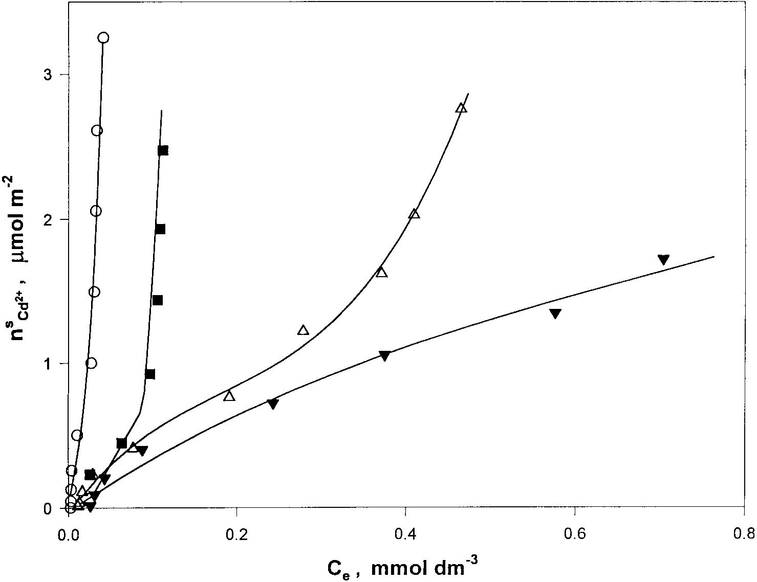
Adsorption excess isotherms of Cd2ϩ ions on HDP-M surfaces (n s, Cd2ϩ) against the concentrations of Cd2ϩ ions present (C e) in ethanol
(1)– cyclohexane (2) mixtures at x ϭ
0.05 (E), 0.1 (■), 0.2 (‚), and 0.4 (Œ).
Adsorption excess isotherms of Cd2ϩ ions on DBS-C surfaces (n s, Cd2ϩ) against the concentrations of Cd2ϩ ions present (C e) in ethanol
(1)– cyclohexane (2) mixtures at x ϭ
0.05 (E), 0.1 (■), 0.2 (‚), and 0.4 (Œ).
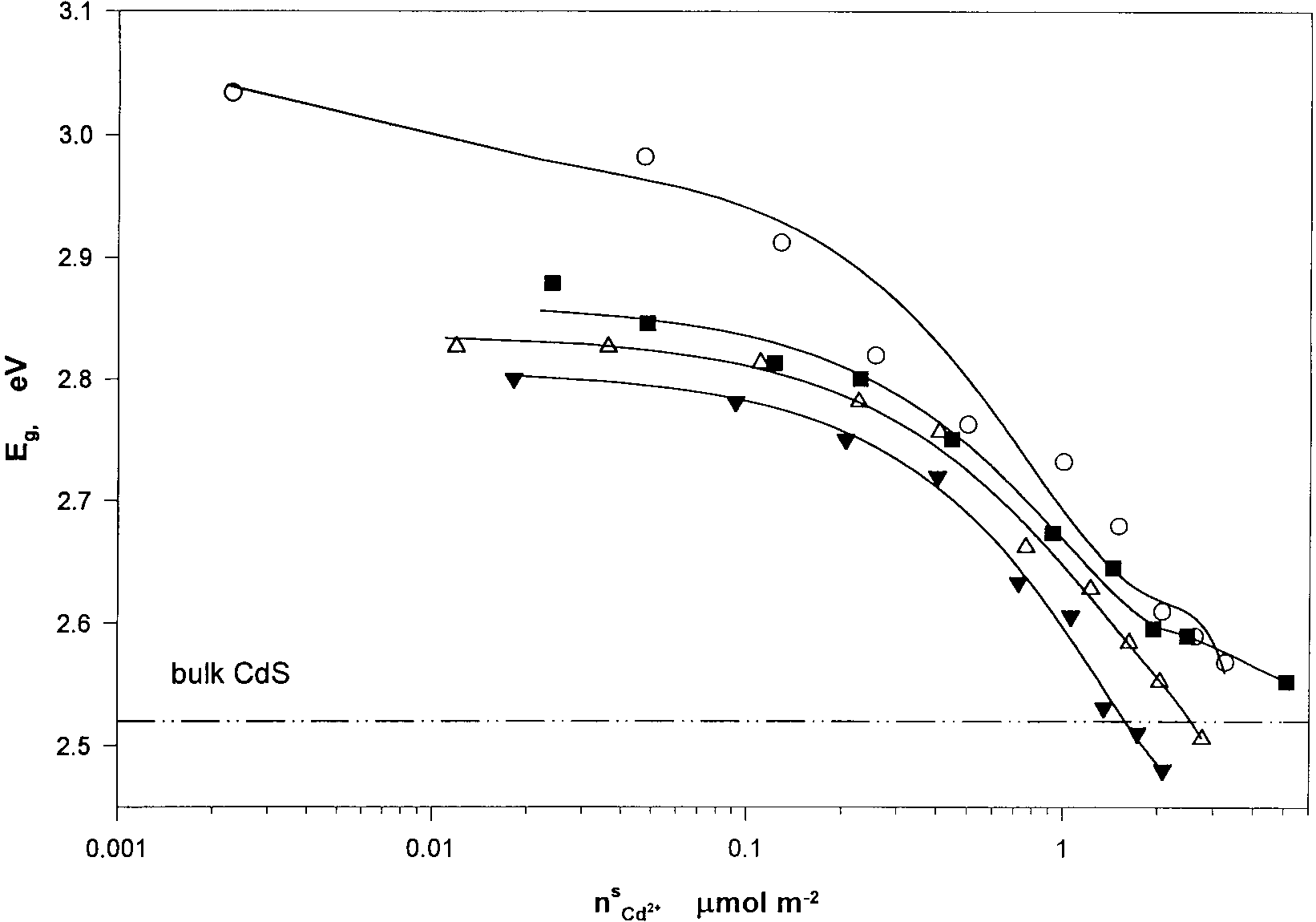
Optical band-gap energies (calculated from the absorption edges) as functions of Cd2ϩ ion adsorbed on HDP-M surfaces in ethanol (1)– cyclohexane
0.05 (E), 0.1 (■), 0.2 (‚), and 0.4 (Œ).
Cd2ϩ ADSORPTION CONTROLS CdS GROWTH IN CLAYS
Optical band-gap energies (calculated from the absorption edges) as functions of Cd2ϩ ion adsorbed on DBS-C surfaces in ethanol (1)– cyclohexane
0.05 (E), 0.1 (■), 0.2 (‚), and 0.4 (Œ).
Absorption spectra were taken on a UVIKON 930 spectro-
Transmission electron microscopic (TEM) images were ob-
tained by means of an OPTON 902 electron microscope,operated at 80 kV. The sizes and size distributions were de-termined by measuring the diameters of 200 –250 particles onphotographic images.
X-ray diffraction measurements were taken on a Phillips PW
1820/1830 diffractometer, (CuK␣) ϭ 0.1542 nm, 50 kV,and 40 mA, in the 1° Յ 2⌰ Յ 32° regime, using the PC-ADP3.5 software. The small-angle diffractions were determined byusing the Cu-␣ line of the PW 1830 generator (50 kV and 40mA) and a KCEC/3 Kratky camera (80 micron diameter,1.5-cm-thick beam). The intensity of the diffracted beam wasdetected by a 100-micron proportional detector using the mov-ing grating method. The intensity of the diffracted beam, I(h),was calculated from
I͑h͒ ϭ I͑h͒
where I(h) and I(h)
sample and the background (empty camera). Here, A ϭ N /N
and A ϭ N /N are the sample and background X-ray dif-
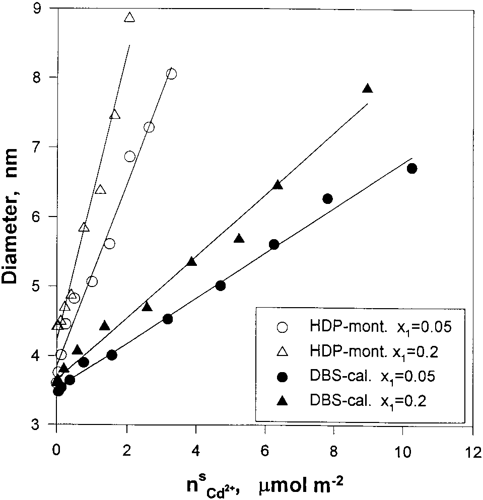
Diameter of CdS particles on HDP-M and DBS-C in various
fraction coefficients, with N , N , and N as the number of
ethanol (1)– cyclohexane (2) mixtures as functions of the adsorption density of
diffracted X-ray photons of the sample, the background, and
the Cd2ϩ cations in ethanol (1)– cyclohexane (2) mixtures at x ϭ
the empty camera, respectively (9 –12).
Diameters of CdS nanoparticles (calculated from the absorption
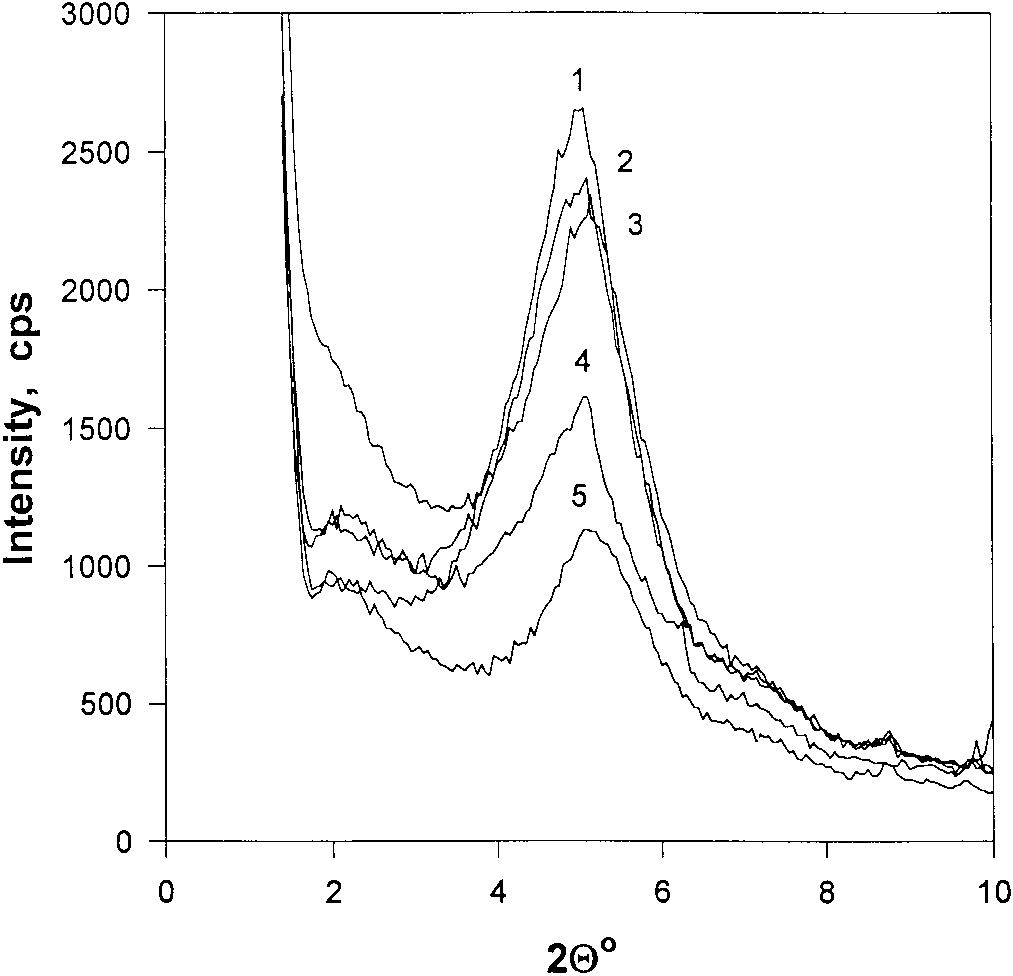
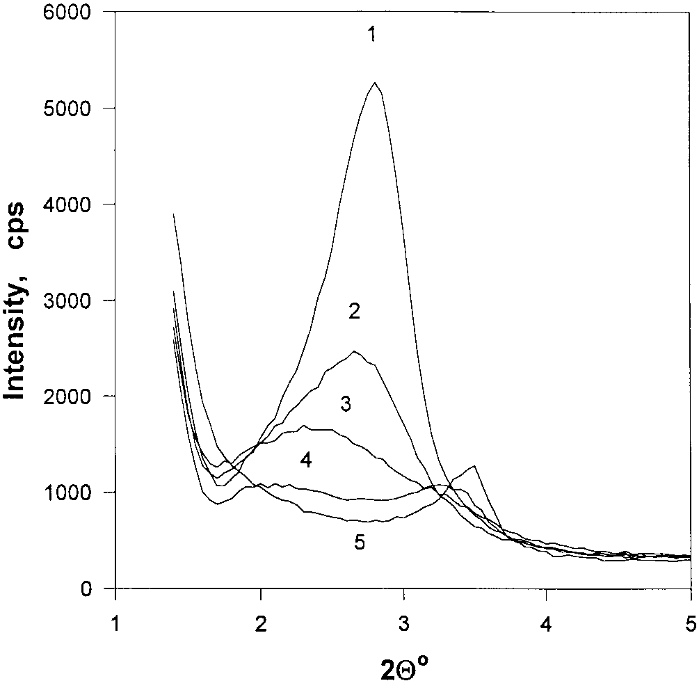
X-ray diffraction patterns of CdS/HDP-M composites of various
edges) intercalated in HDP-M and in DBS-C as functions of excess cadmium
CdS concentrations: (1) 0 mol/g, (2) 4 ϫ 10Ϫ6 mol/g, (3) 4 ϫ 10Ϫ5 mol/g, (4)
ion solubility in ethanol (1)– cyclohexane (2) mixtures at x ϭ
4 ϫ 10Ϫ4 mol/g, (5) 4 ϫ 10Ϫ3 mol/g.
The adsorption isotherms of Cd2ϩ ions onto DBS-C (Fig. 3)
RESULTS AND DISCUSSION
appear to be similar to those determined for HDP-M. Thedifference between the two adsorption excess isotherms (Figs. 1. Cadmiumion Adsorption on Montmorillonite (HDP-M)
2 and 3) is due to the dissimilar surface areas of and adsorption
Cadmium ion concentrations in the ethanolic nanoreactors
have been assessed by adsorption isotherm determinations forthe binding of Cd2ϩ ions onto HDP-M and DBS-C surfaces indifferent ethanol– cyclohexane mixtures.
Cadmium ion solubilities increase, as expected, with in-
creasing mol fractions of ethanol in the ethanol– cyclohexaneliquid mixture (see Fig. 1). Plotted in Fig. 2 are the determinedadsorption excess isotherms of cadmium ions adsorbed onHDP-M (n s
) against the concentrations of Cd2ϩ ion present
(C ) in ethanol (1)– cyclohexane (2) mixtures at x ϭ 0.05,
0.1, 0.2, and 0.4. The amount of cadmium ion adsorbed isseen to depend strongly on the composition of the binaryliquid. Thus, at x ϭ 0.05, 95% of the cadmium ions are
adsorbed on the HDP-M surface; the solubility of Cd2ϩ ion inthe x
ϭ 0.05 ethanol (1)–cyclohexane (2) mixture is 0.5
mmol/dm3 (Fig. 1) and the concentration of Cd2ϩ adsorbed is95% (Fig. 2). The extremely rapid rise of the Cd2ϩ ionsadsorbed at x ϭ 0.05 unequivocally indicates the partitioning
of essentially all of the cadmium ions onto the HDP-M surface(i.e., C Ϸ 0). Increasing the amount of ethanol in the ethanol
cyclohexane mixture resulted in a progressive decrease of theslopes of the adsorption isotherms, and at x
X-ray diffraction patterns of CdS/DBS-C composites of various
surfaces of the HDP-M platelets were found to be essentially
CdS concentrations: (1) 0 mol/g, (2) 4 ϫ 10Ϫ6 mol/g, (3) 4 ϫ 10Ϫ5 mol/g, (4)
4 ϫ 10Ϫ4 mol/g, (5) 4 ϫ 10Ϫ3 mol/g.
Cd2ϩ ADSORPTION CONTROLS CdS GROWTH IN CLAYS
Schematics showing the expansion of the HDP-M (or DBS-C)
lamellas upon the formation of CdS nanoparticles.
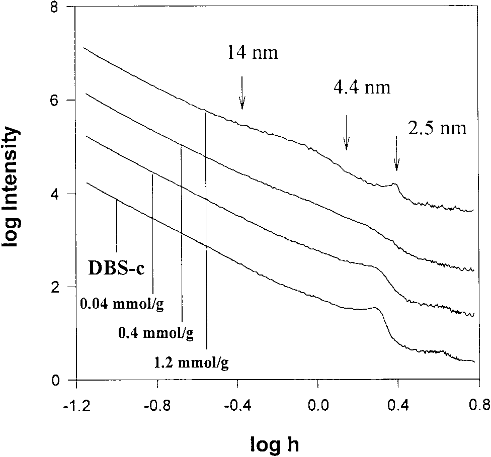
Plots of logarithms of the intensity of the scattered radiation
against the logarithms of the scattering vector (h ϭ 4 sin ⌰/) for CdS/DBS-C composites for various CdS concentrations. The arrows indicate thepoints of d
The volume of the adsorption layer (V s) on a given HDP-M
L values (corresponding to the appropriate 2⌰ values). The curves
are offset by 0.5, 1.0, 1.5, and 2.0 units on the Y-axis.
or DBS-C surface can be calculated by the adsorption spacefilling model (13–15),
components 1 (ethanol) and 2 (cyclohexane), n s is the amount
of adsorbed ethanol, and n s is the adsorbed amount of cyclo-
hexane on the surface of the clay nanoplatelet. If preferentialadsorption of ethanol occurs, then V s ϭ n sV
where n s is the adsorption capacity of the pure ethanol in the
SAXS and BET Data for the Surface Areas of HDP-M and DBS-C in the Presence and in the Absence of CdS Nanoparticles a Kp, tail-end constant in the porod equation.
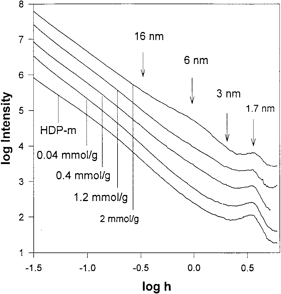
Plots of logarithms of the intensity of the scattered radiation
b S/V, specific surface area of the lamellas relative to their unit volumes.
against the logarithms of the scattering vector (h ϭ 4 sin ⌰/) for CdS/
c Sp, specific surface area of the lamellas relative to their unit mass.
HDP-M composites for various CdS concentrations. The arrows indicate the
d a sBET(N2), specific surface area of the lamellas, determined by BET.
L values (corresponding to the appropriate 2⌰ values). The curves
are offset by 0.5, 1.0, 1.5, and 2.0 units on the Y-axis.
(a) Transmission electron micrograph images of CdS nanoparticles, prepared by the infusion of H2S to 0.8 mmol Cd2ϩ per gram of HDP-M in
ethanol (1)– cyclohexane mixture at x ϭ
Determination of the adsorption excess isotherm, n (n) ϭ
resulted in the formation of CdS particles with a mean diameter
f( x ), as reported previously (14 –16), led to the values n s ϭ
of 3.6 nm. Increasing x to 0.4 in the same system resulted in
4.75 mmol/g, n s ϭ 3.59 mmol/g, and n s ϭ 11.0 mmol/g for
the formation of CdS particles with a mean diameter of 4.9 nm.
HPD-M. For DBS-C, n s ϭ 1.05 mmol/g, n s ϭ 0.91 mmol/g,
A similar tendency was observed on using DBS-C surfaces.
and n s ϭ 2.71 mmol/g, using the Schay–Nagy extrapolation
The relationship between the diameters (d) of the incipient
method (15), or by the Everett–Schay function (13). These
CdS nanoparticles and the concentration of the cadmium ions
values, in turn, permitted the calculation of the volume of the
(Q, in mmol/dm3) can also be illustrated by plotting d against
adsorption layer (i.e., the volume of the nanoreactors, V s) to be
0.664 cm3/g for HPD-M and 0.164 cm3/g for DBS-C.
diameter of the CdS particles formed can also be plottedagainst the excess cadmium ion solubility, (Q Ϫ L)/L (where
2. Absorption Spectra of CdS Nanoparticles GeneratedL is the solubility of CdS in the liquid mixture, taken to be
in Situ on Montmorillonite and Calumit Surfaces
8.9 ϫ 10Ϫ8/dm3). Plots of d vs (Q Ϫ L)/L in HPD-M and in
Absorption spectra of CdS dispersions, in situ generated on
DBS-C (Fig. 7) also illustrate that the smallest nanoparticles
HPD-M and DBS-C surfaces, indicated the size quantization of
are formed in the liquid mixture which contains the least
the nanoparticles. Indeed, the observed absorption edges per-
mitted the assessment of the optical band-gap (E ) and the
diameter of the semiconductor particles by the Brus equation
3. XRD, SAXS, and TEM of CdS Nanoparticles Intercalated
(5– 8, 17, 18). The optical bandgaps of the CdS nanoparticles,
into Layered Montmorillonite and Calumit ClaysE , were found to correlate well with the amount of cadmium
ions adsorbed (Figs. 4 and 5). As can be seen, the smallest CdS
The X-ray diffraction pattern of HPD-M (in solid powder
particles are formed in the ethanol– cyclohexane mixture which
samples) is characterized by a broad peak at 2⌰ ϭ (5.0) or dL
contains the least amount of ethanol (i.e., x ϭ 0.05). For
ϭ 1.76 nm (curve 1, Fig. 8) which corresponds to the basal
example, incorporation of 0.02 mol of cadmium ions into the
spacing of the hydrophobic montmorillonite sheets. The posi-
ethanolic x ϭ 0.05 nanoreactor on the surface of HPD-M
tion of this peak has been shown to depend on the extent of
Cd2ϩ ADSORPTION CONTROLS CdS GROWTH IN CLAYS
CdS intercalation in HPD-M. With increased CdS particle
Transmission electron microscopic investigations estab-
intercalation the intensity of the XRD peaks decreased and
lished the formation of relatively monodispersed CdS particles
shifted to larger 2⌰ values (curve 2, 2⌰ ϭ 4.8, d ϭ 1.84 nm;
curve 3, 2⌰ ϭ 5.2, d ϭ 1.69 nm; curve 4, 2⌰ ϭ 5.2, d ϭ
1.69 nm; and 2⌰ ϭ 2.05, d ϭ 4.31 nm; curve 5, 2⌰ ϭ 2.0,
CONCLUSIONS
Information on the selective adsorption of cadmium ions
The X-ray diffraction pattern of DBS-C samples is quite
into polar nanoreactors, formed on montmorillonite and calu-
similar. It is characterized by a high-intensity broad peak at 2⌰
mit surfaces in binary ethanol– cyclohexane mixtures, has been
2.75° or d ϭ 3.21 nm (curve 1, Fig. 9), which corresponds
obtained in the present work by adsorption isotherm determi-
to the basal spacing in intercalated DBS calumit sheets (16,
nations. This information has provided, in turn, an additional
19). Intercalation of increasing amounts of CdS nanoparticles
means to control the sizes of semiconductor nanoparticles in
into the DBS-C sheets also resulted in a progressive decrease
situ generated within the lamellas of silica sheets. X-ray dif-
of the XRD peak intensity and a concomitant shift to larger 2⌰
fraction measurements indicated the controllable stretching of
values (curve 2, 2⌰ ϭ 2.6, d ϭ 3.39 nm; curve 3, 2⌰ ϭ 2.4,
the silica sheets by the amount of CdS nanoparticles generated. d ϭ 3.68 nm; and 2⌰ ϭ 2.05, d ϭ 4.31 nm).
This, in turn, opens the door to the organization of nanopar-
The progressive decrease of the intensity and concomitant
ticles into two-dimensional arrays and three-dimensional net-
broadening of the diffraction peaks upon CdS intercalation into
works on silica templates by the juidicious manipulation of
the HPD-M and DBS-C sheets indicate an uneven stretching of
the lamellas upon the formation of CdS nanoparticles and adecrease of their uniformity, as schematically illustrated inFig. 10. ACKNOWLEDGMENTS
Fruitful structural information has been acquired by analyz-
The authors are grateful for the financial support of this work by the U.S.
ing the obtained low-angle X-ray scattering data. In Figs. 11
National Science Foundation and the Hungarian Academy of Science (NSF-
and 12, intensities of the scattered radiation against the scat-
MTA project 84 and AKP 97-141 2,4/18).
tering vector (h ϭ 4 sin ⌰/) are plotted logarithmically forHDP-M and DBS-C in the absence and in the presence of
REFERENCES
intercalated CdS nanoparticles. Progressive broadening of the
1. Enea, O., and Bard, A., J. Phys. Chem. 90, 301 (1986).
of the Bragg peaks (1.7 nm for HDP-M and 2.5 nm for DBS-C)
2. Lianos, P., and Thomas, J. K., Chem. Phys. Lett. 125, 299 –302 (1986).
with increasing amounts of CdS intercalation are also seen in
3. Resch, U., Eychmu¨ller, A., Hasse, M., and Weller, H., Langmuir 8,
Figs. 11 and 12. Taking advantage of the Porod plot (i.e.,
plotting Ih 3 vs h 3, not shown), we obtained the the tail-end
4. Vossmeyer, T., Katsikas, L., Giersig, M., Popovic, I. G., Desner, K.,
Chemseddine, A., Eychmu¨ller, A., and Weller, H., J. Phys. Chem. 98,
(as h 3 3 ϱ). Knowledge of K permits the
calculation of the specific surface area of the lamellas relative
5. Kotov, N. A., Putyera, K., Fendler, J. H., Tomba´cz, E., and De´ka´ny, I.,
to their unit volume, S/V from (10, 11)
Colloids Surf. A 71, 317–326 (1993).
6. De´ka´ny, I., Turi, L., Tomba´cz, E., and Fendler, J. H., Langmuir 11,
2285–2292 (1995); Rossetti, R., Hull, R., Gibson, J. M., and Brus, L. E.,
J. Phys. Chem. 82, 552–559 (1985)
7. De´ka´ny, I., Nagy, L., Turi, L., Kira´ly, Z., Kotov, N. A., and Fendler, J. H.,
where and are the volume fractions of solids and holes,
Langmuir 12, 3709 –3715 (1996); Kamat, P. V., and Dimitrijevic, N. M.,
respectively, and M is the Porod invariant (10, 11). Further-
J. Phys. Chem. 98, 7665 (1994).
more, knowing the apparent density of the solid powder in the
8. De´ka´ny, I., Turi, L., Galba´cs, G., and Fendler, J. H., J. Colloid InterfaceSci. 195, 307–315 (1997).
sample holder, d, we can calculate the specific surface area of
9. Guinier, A., and Fournet, G., “Small Angle Scattering of X-Rays.” Wiley,
the interior surfaces relative to the unit mass:
10. Glatter, O., and Kratky, O., “Small-Angle X-Ray Scattering.” Academic
11. Porod, G., Kolloid Z. 124, 83 (1951); Porod, G., Kolloid Z. 125, 1 (1952). 12. Stabinger, H., and Kratky, O., Macromol. Chem. 179, 1655 (1978).
Values of K , S/V, and S for HDP-M and DBS-C in the
13. Schay, G., and Nagy, L. Gy., J. Colloid Interface Sci. 38, 302 (1972).
absence and in the presence of intercalated CdS nanoparticles
14. De´ka´ny, I., Sza´nto´, F., and Nagy, L. G., J. Colloid Interface Sci. 109, 376 (1986).
are collected in Table 1. Table 1 also reports the specific areas
15. De´ka´ny, I., and Nagy, L. G., J. Colloid Interface Sci. 147, 119 (1991).
available for gas adsorption in HDP-M, determined by the BET
16. De´ka´ny, I., Berger, F., Imrik, K., and Lagaly, G., Colloid Polym. Sci. 275,
method. The specific surface areas of the lamellas, expressed
17. Brus, L. E., J. Chem. Phys. 79, 5566 –5571 (1983).
either in terms of S/V or as S , are seen to increase with
18. Brus, L. E., J. Chem. Phys. 80, 4403– 4409 (1984).
increasing amounts of CdS incorporated.
19. Meyn, M., Beneke, K., and Lagaly, L., Inorg. Chem. 29, 5201 (1990).
Dermatologie L’incidence de la gale a augmenté de 10 % en France en 10 ans confirmant une tendance observée à l’étranger et soulignant les difficultés thérapeutiques. Le rapport du Haut Comité de la santé publique conseille, dans un souci de simplification des protocoles, un 1er traitement à J1 et un second, systématiquement, une semaine après. LA PRISE EN CHARGE DE LA G

 Journal of Colloid and Interface Science 213, 584 –591 (1999)
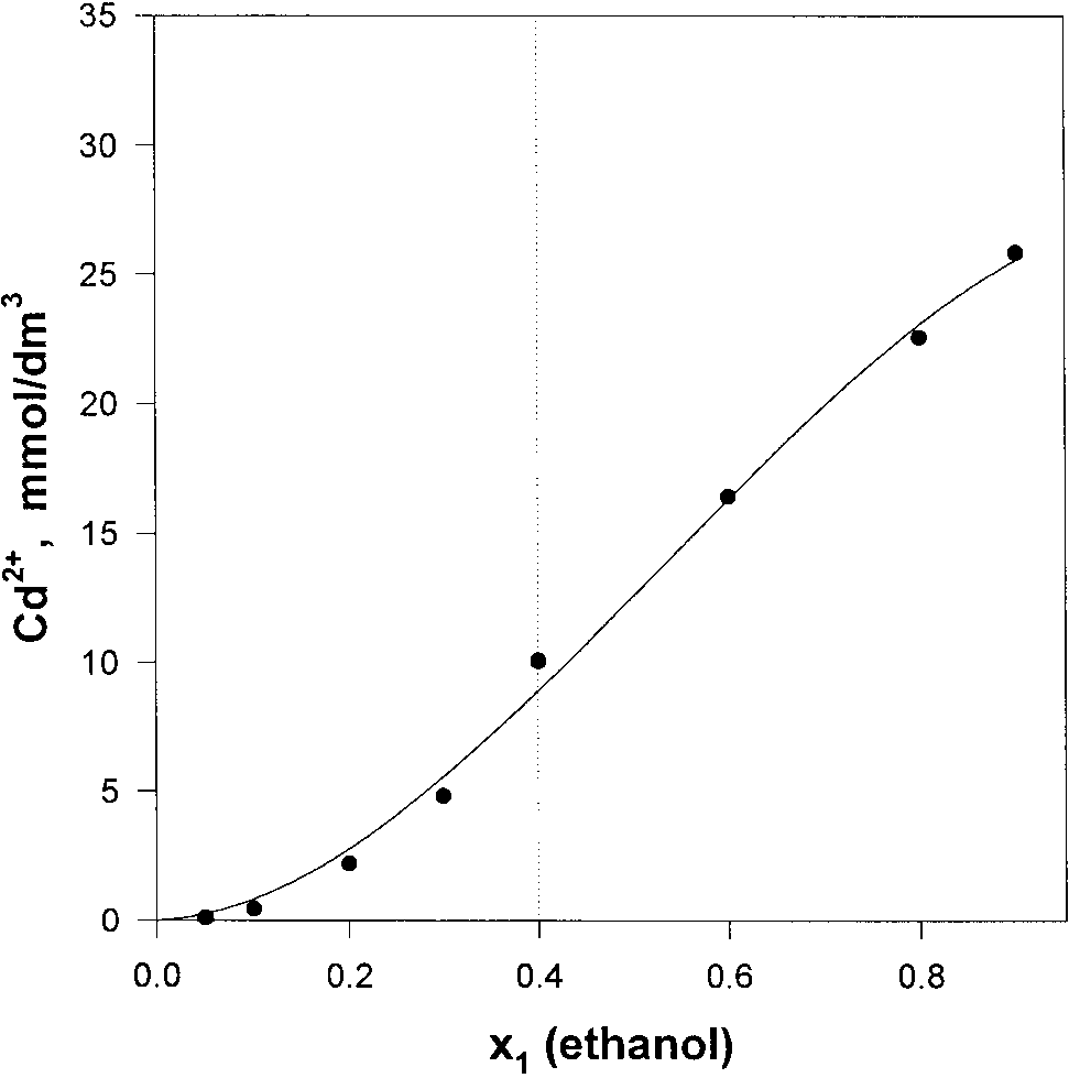
 Cd2ϩ ADSORPTION CONTROLS CdS GROWTH IN CLAYS
The sample solutions were introduced to the nebulizer of the
ICP-AES spectrometer by a high-precision Gilson MinipulseIII peristaltic pump at a flow rate of 1.5 cm3/min. The argonplasma spectrometer was operated at 1 kW plasma forwardpower and 12 L/min plasma gas and 1 L/min sheath gas flowrate settings. Three-point linear calibration was performed,covering the range of concentration of interest, 1–100 ppm.
Cd2ϩ ADSORPTION CONTROLS CdS GROWTH IN CLAYS
The sample solutions were introduced to the nebulizer of the
ICP-AES spectrometer by a high-precision Gilson MinipulseIII peristaltic pump at a flow rate of 1.5 cm3/min. The argonplasma spectrometer was operated at 1 kW plasma forwardpower and 12 L/min plasma gas and 1 L/min sheath gas flowrate settings. Three-point linear calibration was performed,covering the range of concentration of interest, 1–100 ppm.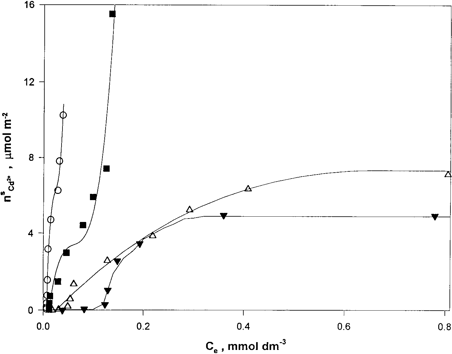
 Adsorption excess isotherms of Cd2ϩ ions on DBS-C surfaces (n s, Cd2ϩ) against the concentrations of Cd2ϩ ions present (C e) in ethanol
(1)– cyclohexane (2) mixtures at x ϭ
0.05 (E), 0.1 (■), 0.2 (‚), and 0.4 (Œ).
Adsorption excess isotherms of Cd2ϩ ions on DBS-C surfaces (n s, Cd2ϩ) against the concentrations of Cd2ϩ ions present (C e) in ethanol
(1)– cyclohexane (2) mixtures at x ϭ
0.05 (E), 0.1 (■), 0.2 (‚), and 0.4 (Œ).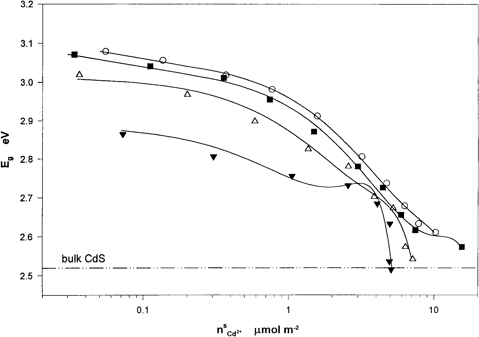
 Cd2ϩ ADSORPTION CONTROLS CdS GROWTH IN CLAYS
Optical band-gap energies (calculated from the absorption edges) as functions of Cd2ϩ ion adsorbed on DBS-C surfaces in ethanol (1)– cyclohexane
0.05 (E), 0.1 (■), 0.2 (‚), and 0.4 (Œ).
Cd2ϩ ADSORPTION CONTROLS CdS GROWTH IN CLAYS
Optical band-gap energies (calculated from the absorption edges) as functions of Cd2ϩ ion adsorbed on DBS-C surfaces in ethanol (1)– cyclohexane
0.05 (E), 0.1 (■), 0.2 (‚), and 0.4 (Œ).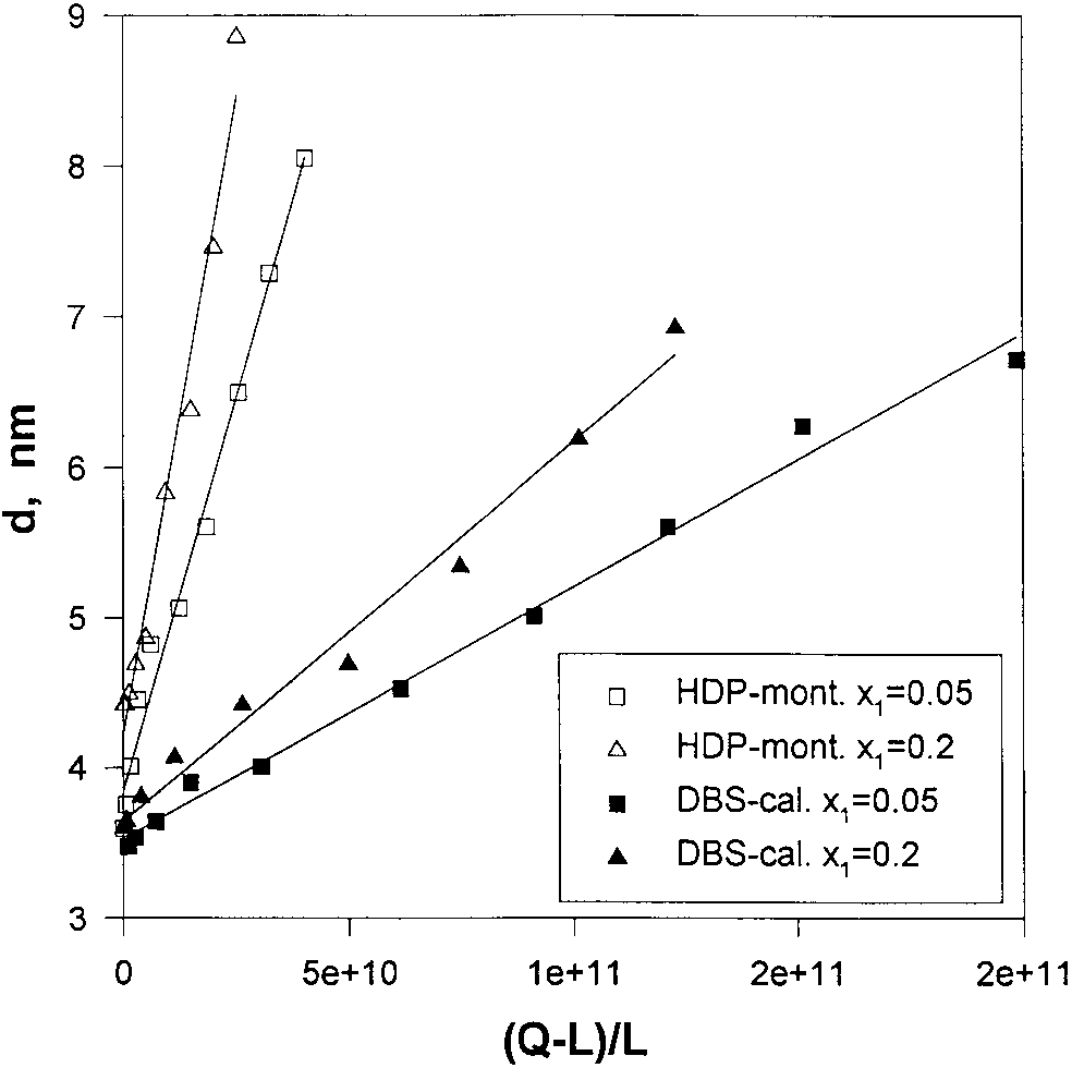
 Diameters of CdS nanoparticles (calculated from the absorption
X-ray diffraction patterns of CdS/HDP-M composites of various
edges) intercalated in HDP-M and in DBS-C as functions of excess cadmium
CdS concentrations: (1) 0 mol/g, (2) 4 ϫ 10Ϫ6 mol/g, (3) 4 ϫ 10Ϫ5 mol/g, (4)
ion solubility in ethanol (1)– cyclohexane (2) mixtures at x ϭ
4 ϫ 10Ϫ4 mol/g, (5) 4 ϫ 10Ϫ3 mol/g.
Diameters of CdS nanoparticles (calculated from the absorption
X-ray diffraction patterns of CdS/HDP-M composites of various
edges) intercalated in HDP-M and in DBS-C as functions of excess cadmium
CdS concentrations: (1) 0 mol/g, (2) 4 ϫ 10Ϫ6 mol/g, (3) 4 ϫ 10Ϫ5 mol/g, (4)
ion solubility in ethanol (1)– cyclohexane (2) mixtures at x ϭ
4 ϫ 10Ϫ4 mol/g, (5) 4 ϫ 10Ϫ3 mol/g.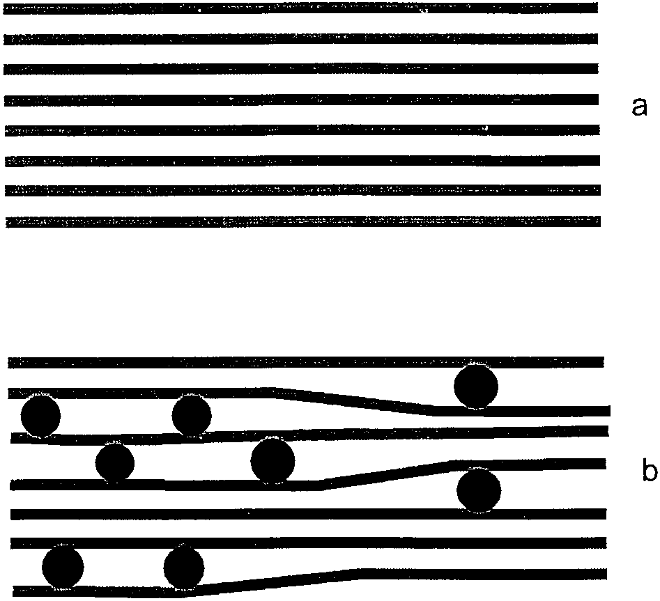
 Cd2ϩ ADSORPTION CONTROLS CdS GROWTH IN CLAYS
Schematics showing the expansion of the HDP-M (or DBS-C)
lamellas upon the formation of CdS nanoparticles.
Cd2ϩ ADSORPTION CONTROLS CdS GROWTH IN CLAYS
Schematics showing the expansion of the HDP-M (or DBS-C)
lamellas upon the formation of CdS nanoparticles.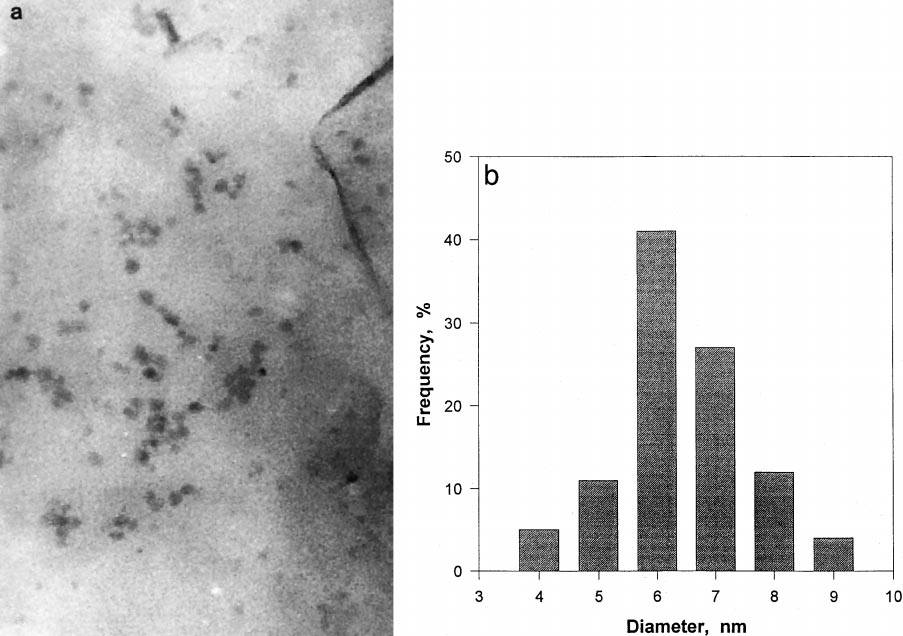 (a) Transmission electron micrograph images of CdS nanoparticles, prepared by the infusion of H2S to 0.8 mmol Cd2ϩ per gram of HDP-M in
ethanol (1)– cyclohexane mixture at x ϭ
Determination of the adsorption excess isotherm, n (n) ϭ
resulted in the formation of CdS particles with a mean diameter
f( x ), as reported previously (14 –16), led to the values n s ϭ
of 3.6 nm. Increasing x to 0.4 in the same system resulted in
4.75 mmol/g, n s ϭ 3.59 mmol/g, and n s ϭ 11.0 mmol/g for
the formation of CdS particles with a mean diameter of 4.9 nm.
(a) Transmission electron micrograph images of CdS nanoparticles, prepared by the infusion of H2S to 0.8 mmol Cd2ϩ per gram of HDP-M in
ethanol (1)– cyclohexane mixture at x ϭ
Determination of the adsorption excess isotherm, n (n) ϭ
resulted in the formation of CdS particles with a mean diameter
f( x ), as reported previously (14 –16), led to the values n s ϭ
of 3.6 nm. Increasing x to 0.4 in the same system resulted in
4.75 mmol/g, n s ϭ 3.59 mmol/g, and n s ϭ 11.0 mmol/g for
the formation of CdS particles with a mean diameter of 4.9 nm.