Gene gun bombardment with dna-coated gold particles is a potential alternative to hydrodynamics-based transfection for delivering genes into superficial hepatocytes
HUMAN GENE THERAPY 19:391–395 (April 2008) Mary Ann Liebert, Inc. DOI: 10.1089/hum.2007.152 Technical Report
Gene Gun Bombardment with DNA-Coated Gold Particles Is
a Potential Alternative to Hydrodynamics-Based Transfection
for Delivering Genes into Superficial Hepatocytes
MING-LING CHANG,1 JENG-CHANG CHEN,2 CHAU-TING YEH,1 MING-YU CHANG,3
CHUN-KAI LIANG,1 CHENG-TANG CHIU,1 DENG-YN LIN,1 and YUN-FAN LIAW1
ABSTRACT Although in vivo nonviral gene delivery to the liver is critical for hepatic gene therapy, there are a number of technical obstacles. Enhanced green fluorescent protein (EGFP)-encoding DNA was coated onto gold par- ticles (gold–DNA), dissolved in phosphate-buffered saline (pure DNA), and prepared as a polymer adjuvant (jetPEI)–galactosidase solution (polymer–DNA). Murine liver transfection was attempted by nonviral ap- proaches, which included hydrodynamics-based transfection (HBT) of pure DNA, transport and transhepatic injection of polymer–DNA, and gene gun bombardment with pure DNA, gold–DNA, and polymer–DNA. Only HBT and gene gun bombardment yielded significant numbers of EGFPϩ hepatocytes. With the exception of the edge of the liver, HBT had a whole-liver transfection rate of 20% under optimized conditions. HBT re- sulted in marked hepatic infarctions, most prominently at the edge of the liver. For gene gun bombardment, the transfection rate was pressure dependent and limited to 15% for gold–DNA. Triple or quadruple bom- bardment at 30 psi resulted in a transfection rate comparable to that of a single bombardment at higher pres- sure, but was associated with minimal scattered hepatic necrosis. The EGFPϩ hepatocytes were located mainly in the superficial layers. We conclude that both HBT and gene gun bombardment yielded efficient murine hepatocyte transfection in vivo. Severe hepatic infarction impedes foreign gene expression in the superficial hepatocytes after HBT. Repeated bombardment with gold–DNA, using an accelerated particle gene gun at 30 psi, is a potential alternative to HBT for delivering genes to superficial hepatocytes in vivo, although gold-re- lated hepatic necrosis is a persistent problem. INTRODUCTION
attractive because it can be manipulated by standard recombi-nant DNA techniques and delivered by both chemical and phys-
IN VIVO gene delivery to the liver is critical for both experi- ical means. However, chemical approaches such as circulating
mental and clinical applications. At present, there are two
cationic vectors can attract serum proteins, leading to dynamic
main modes for gene delivery: viral and nonviral (Dobson,
changes in their physicochemical properties and diminished
2006). Viral vectors confer more effective expression than syn-
transfection efficiency (Nishikawa and Huang, 2001). Physical
thetic molecular gene vectors, albeit at the expense of infection
approaches to gene transfer have improved and become as ef-
and immunogenicity (Azzam and Domb, 2004). To lessen the
fective as viral vectors (Wells, 2004). Hydrodynamics-based
potential biohazards of viral vectors, naked DNA is considered
transfection (HBT) of hepatocytes has been reported to produce
1Liver Research Center and Department of Hepatogastroenterology, Chang Gung Memorial Hospital, Taoyuan, Taiwan; and Chang Gung Uni-
2Department of Surgery, Chang Gung Children’s Hospital, Taoyuan, Taiwan 33305. 3Division of Pediatric Critical Care and Emergency Medicine, Chang Gung Children’s Hospital, Taoyuan, Taiwan 33305. CHANG ET AL.
a satisfactory transfection efficiency in mice (Wolff and Bud-
vivo transfection via the portal vein (100 to 400 l for 10 min)
ker, 2005). Notably, gene guns can be used for difficult-to-trans-
or direct injected into the right lobe of the liver (20 to 100 l
fect cells and particular in situ approaches (Johnston and Tang,
for 3 min). Tail vein injection was also performed (400 l for
1994). However, whether gene guns are effective for liver trans-
fection is uncertain. We examined the effectiveness of murineliver transfection by gene gun bombardment with enhanced
green fluorescent protein (EGFP)-encoding DNA and compared
Five to 250 g of EGFP DNA was injected via the tail vein
the results with those obtained by other chemical or physical
in a volume of saline equivalent to 8% of the body mass of the
mouse (e.g., 1.6 ml for a 20-g mouse). The entire volume wasdelivered within 5 sec. MATERIALS AND METHODS
Mice were killed 48 hr or 7 days after transfection, and their
livers were harvested. The livers were either cryofixed or fixed
Eight-week-old male FVB/N mice were purchased from the
in 4% buffered paraformaldehyde (PFA). Unless otherwise in-
Animal Center of the National Science Council (Taipei, Tai-
dicated, transfection rates were evaluated 48 hr after transfec-
wan). For each transfection method, 30 mice were used. The
use of animals in this study was approved by the Animal Care
Cryofixation was performed by immersion of tissues in ice-
and Use Committee at Chang Gung Memorial Medical Center
cold isopentane for 3 min, followed by freezing at -80°C. Fixed
frozen samples were mounted in Tissue-Tek O.C.T. 4583 com-
pound (Sakura Finetek USA, Torrance, CA). Samples were sec-tioned sequentially on a Jung Frigocut 2800N (Leica, Deerfield,
EGFP plasmid (PEGFP-C1, 4.7 kDa) was purchased from
IL) at a cutting interval of 6 m. Samples fixed in 4% PFA
Clontech (Mountain View, CA). The plasmid was cloned and
were subjected to hematoxylin and eosin (H&E) staining. Sec-
purified with an EndoFree plasmid kit (Qiagen, Valencia, CA).
tions were examined by either fluorescence microscopy or light
Naked EGFP DNA was dissolved at 1 g/l in phosphate-
microscopy. EGFPϩ hepatocytes were observed at ϫ20 mag-
buffered saline (PBS) (pure DNA). EGFP DNA-coated gold
nification under the fluorescence microscope. The transfection
particles (gold–DNA) were prepared by adding 5 mg of Bi-
rate was defined as the number of EGFPϩ hepatocytes divided
olistic 1.0-m gold particles (Bio-Rad, Hercules, CA) to 5 l
by the total number of hepatocytes within the same field on
of 1-g/l plasmid solution, 20 l of 0.1 M spermidine (Sigma-
three randomized occasions. Mice transfected with DNA-free
Aldrich, St. Louis, MO), and 20 l of 0.5 M CaCl2 (Sigma-
PBS (with or without gold) of the same volume were used as
Aldrich). After several washes, the precipitate was dissolved in
100% alcohol for bombardment. The EGFP DNA–jetPEI–Galsolution (polymer–DNA) was prepared according to the man-
ufacturer’s protocol (Polyplus Transfection, New York, NY).
Forty-eight hours after transfection, the serum alanine amino-
The ratio of nitrogen residues on jetPEI to phosphates on the
transferase (ALT) levels of the mice were measured with a
DNA backbone (N:P ratio) ranged from 5 to 10 for 0.31 to 0.62
Vitros DT60 II chemistry system (Johnson & Johnson, New
Gene gun transfection with pure DNA, gold–DNA,
Independent sample t testing was used to compare the means
After general anesthesia by intraperitoneal injection of ket-
obtained for two different bombardment pressures or repeti-
amine and diphenhydramine (Benadryl; Pfizer, New York, NY),
tions. One-way analysis of variance (ANOVA) was used to test
the mice underwent midline laparotomy, to exposure the liver
the equality of the means among the three DNA groups. Dif-
for gene gun bombardment. In situ liver transfections were per-
ferences were regarded as significant for p Ͻ 0.05.
formed with the low pressure-accelerated particle gene gun(Bioware Technologies, Taipei, Taiwan). A 1-cm-thick rubberring was placed on the shooting end of gene gun. Briefly,gold–DNA (5–20 l) was bombarded into mouse liver at pres-
sures of 20–45 psi. Alternatively, pure DNA (5–20 l) wasbombarded into mouse liver at pressures of 20–45 psi. For poly-
Gene gun transfection with pure DNA, gold–DNA, and
mer–DNA bombardment, DNA–jetPEI–Gal solution (5 l) was
bombarded into mouse liver. The mouse abdomen was closed
Mice transfected with the EGFP plasmid by gene gun bom-
bardment did not have significant numbers of EGFPϩ hepato-cytes unless a pressure of 30 psi was used (Figs. 1A and 2). Intravenous or direct liver injection of polymer–DNA
With respect to transfection rate, gold–DNA compared favor-
Mouse liver was exposed as described above. EGFP
ably with pure DNA and polymer–DNA (Fig. 2). However, liver
DNA–jetPEI–Gal solution (N:P ratio, 5–10) was used for in
laceration increased abruptly at pressures above 30 psi, ac-
GENE GUN BOMBARDMENT FOR IN SITU LIVER TRANSFECTION
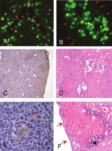
(A and B) EGFPϩ hepatocytes are shown (original magnification, ϫ20) after gene gun bombardment with gold–DNA
(A) at a pressure of 30 psi, and after HBT with injection of 10 g of DNA within 4 sec (B). The edge of the liver is indicated by red arrows. (C and E) H&E staining of mouse liver after gold–DNA bombardment; a low-power field (C, ϫ100) and a high- power field (E, ϫ400) are shown. Gold particles (red arrows) and inflammatory cells (white arrow) are scattered in an area of necrotic hepatocytes (red arrowheads). (D and F) H&E staining of mouse liver after HBT; a low-power field (D, ϫ100) and a high-power field (F, ϫ400) are shown. Diffuse infarctions in the hepatic parenchyma are evident (D). A representative conflu- ent hepatic infarction (F, red arrows) is located underneath the edge of the liver. Infiltrating inflammatory cells (F, white arrows) and calcification (F, black arrow) are associated with the infarction. CHANG ET AL.
were scarce at the edge of the liver (Fig. 1B, arrows). H&Estaining revealed remarkable hepatic infarctions in both controland experimental animals. At the edge of the liver, confluentinfarctions were impressive and formed broad bands (Fig. 1D
and F). The ALT levels of mice were 588 Ϯ 135 U/liter. Oneweek after HBT, the transfection rate decreased to 11.8%. DISCUSSION
The chemical approach with jetPEI-Gal injection in FVB/N
mice was unsatisfactory, as it gave minimal transfection rates
regardless of the injection route. Successful in vivo transfec-
tions by jetPEI injection have been reported in the lung (Zou
et al., 2000). Although jetPEI-Gal was chosen over jetPEI for
use in the current study, because of its higher affinity for he-patocytes (Robaczewska et al., 2001), our data indicate that the
Relationships between the transfection rate and bom-
liver represents a more robust barrier for polymer–adjuvant
bardment pressure for three DNA preparations. In terms of
transfection efficiency, gold–DNA compares favorably with
HBT yielded the highest transfection rate of all the nonviral
pure DNA and polymer–DNA at a pressure of :30 psi (p Ͻ
DNA delivery methods. This is comparable to the results of
0.001, one-way ANOVA). For each DNA preparation, signifi-cant differences were observed for 25 versus 30 psi, 30 versus
previous studies (Zhang et al., 1999; Yang et al., 2002). How-
35 psi, 35 versus 40 psi, and 40 versus 45 psi (p Ͻ 0.001–0.044,
ever, rapid injection of a large volume via the tail vein usually
t test), but not for 40 versus 45 psi in the gold–DNA group (p ϭ
causes transient heart dysfunction (Zhang et al., 2004) and may
lead to animal loss. Clinical application is not feasible, becausehumans lack a homolog for the tail vein. Furthermore, HBTleads to increased venous pressure (Zhang et al., 2004) and sub-
counting for a mortality rate of more than 35%. The maximal
sequent hepatic infarction. The infarctions had a tendency to be
transfection rate achieved by a single bombardment was ap-
confluent at the liver edge, where perfusion is sparser than else-
proximately 15% for gold–DNA and 5–6% for pure DNA or
where. Thus, it does not guarantee foreign gene expression in
polymer–DNA (Fig. 2). At 30 psi, the transfection rates reached
a plateau at approximately 6.2, 5.9, and 15% for pure DNA,polymer–DNA, and gold–DNA, respectively, with three or fourbombardments (Fig. 3). The mortality rate after triple bom-
bardment at 30 psi was negligible and ranged from 0 to 3.3%. Further repetitions of bombardment led to mortality due to gross
liver laceration. Regardless of the composition of the DNA so-
lution, EGFPϩ hepatocytes after bombardment were locatedmainly in the superficial layers (depth of 10–60 m, one to
three cell layers) of the liver. Despite the better transfection rate
obtained for gold–DNA, H&E staining of bombarded liver tis-
sues revealed several necrotic spots with deposition of gold par-
ticles (Fig. 1C and E), indicating liver injury at the bombard-
ment site, probably caused by the gold particles. The ALT levels
of the mice were 249 Ϯ 75 U/liter (normal range, 15–84
U/liter). One week after bombardment with gold–DNA, the
transfection rate decreased to 9.7%. Intravenous and direct liver injection of polymer–DNA
Transfection rates in relation to number of bombard-
None of the transfections with polymer–DNA generated
ment repetitions at a pressure of 30 psi. For the same number
of bombardment repetitions, gold–DNA gives superior trans-fection rates compared with pure DNA and polymer–DNA (p Ͻ
0.001, one-way ANOVA). A significant increase in transfec-tion rate is observed for bombardment performed up to three
The immediate mortality rate was 6.6%, despite cardiopul-
times for each DNA group (p Ͻ 0.001–0.032, t test), with the
monary resuscitation for more than 10 min. The highest trans-
exception of polymer–DNA bombardment carried out once and
fection rate for HBT was about 20% under optimized condi-
twice (p ϭ 0.075, t test). The transfection rates for three and
tions of :10 g of DNA injected within 4 sec. EGFPϩ
four bombardment repetitions are not significantly different for
hepatocytes were evenly distributed over the whole liver but
each group (p ϭ 0.55–1.0, t test). GENE GUN BOMBARDMENT FOR IN SITU LIVER TRANSFECTION
The original application of the gene gun was for skin vacci-
REFERENCES
nation, which induces DNA expression in the most superficiallayers of the skin (Johnston and Tang, 1994; Peachman et al.,
AZZAM, T., and DOMB, A.J. (2004). Current developments in gene
2003). Thus, cell sampling for gene gun bombardment should
transfection agents. Curr. Drug Deliv. 1, 165–193.
focus on the superficial cells. GFPϩ hepatocytes were most
DOBSON, J. (2006). Gene therapy progress and prospects: Magnetic
prominent in the first three layers. In comparison with the skin,
nanoparticle-based gene delivery. Gene Ther. 13, 283–287.
the liver is too fragile to bear the bombardment pressure re-
JOHNSTON, S.A., and TANG, D.C. (1994). Gene gun transfection of
animal cells and genetic immunization. Methods Cell Biol. 43,
quired for in situ transfection. Therefore, the pressure must be
adjusted by weighing transfection efficiency against possible
NISHIKAWA, M., and HUANG, L. (2001). Nonviral vectors in the
liver tearing. Triple bombardment at a tolerable pressure of 30
new millennium: Delivery barriers in gene transfer. Hum. Gene Ther.
psi has been shown to yield a transfection efficiency compara-
12, 861–870.
ble to that obtained from a single bombardment at higher pres-
PEACHMAN, K.K., RAO, M., and ALVING, C.R. (2003). Immu-
sure, which usually causes gross liver laceration. However, un-
nization with DNA through the skin. Methods 31, 232–242.
predictable location of gene transfer usually ensues from a
ROBACZEWSKA, M., GUERRET, S., REMY, J.S., CHEMIN, I.,
direct strike (our unpublished data). Therefore, a rubber ring
OFFENSPERGER, W.B., CHEVALLIER, M., BEHR, J.P., POD-
was placed at the opening of the gene gun. It ensures good guid-
HAJSKA, A.J., BLUM, H.E., TREPO, C., and COVA, L. (2001).
ance, allowing constant focusing and an attenuated blast effect.
Inhibition of hepadnaviral replication by polyethylenimine-based intravenous delivery of antisense phosphodiester oligodeoxynu-
Among the various DNA preparations, gold–DNA bombard-
cleotides to the liver. Gene Ther. 8, 874–881.
ment had the highest transfection rate, although it was associ-
WELLS, D.J. (2004). Gene therapy progress and prospects: electropo-
ated with gold particle-related necrosis. Nevertheless, the level
ration and other physical methods. Gene Ther. 11, 1363–1366.
of injury, determined by histological examination and the serum
WOLFF, J.A., and BUDKER, V. (2005). The mechanism of naked
ALT level, was less severe than that caused by HBT. Gene gun
DNA uptake and expression. Adv. Genet. 54, 3–20.
bombardment is comparable with HBT in terms of stability,
YANG, P.L., ALTHAGE, A., CHUNG, J., and CHISARI, F.V. (2002).
with a 20% decrease in the transfection rate after 1 week.
Hydrodynamic injection of viral DNA: A mouse model of acute he-
In conclusion, gene gun bombardment of the liver with
patitis B virus infection. Proc. Natl. Acad. Sci. U.S.A. 99, 13825–13830.
gold–DNA is a potentially useful alternative to HBT for the
ZHANG, G., BUDKER, V., and WOLFF, J.A. (1999). High levels of
transfection of superficial hepatocytes, particularly because
foreign gene expression in hepatocytes after tail vein injections of naked plasmid DNA. Hum. Gene Ther. 10, 1735–1737.
it does not induce severe hepatic injury. Its application could
ZHANG, G., GAO, X., SONG, Y.K., VOLLMER, R., STOLZ, D.B.,
potentially be extended to other animals, regardless of the
GASIOROWSKI, J.Z., DEAN, D.A., and LIU, D. (2004). Hy-
presence or absence of a tail vein; however, its application is
droporation as the mechanism of hydrodynamic delivery. Gene Ther.
limited to superficial cells and animals that can tolerate lap-
11, 675–682.
ZOU, S.M., ERBACHER, P., REMY, J.S., and BEHR, J.P. (2000). Sys-
temic linear polyethylenimine (L-PEI)-mediated gene delivery in the mouse. J. Gene Med. 2, 128–134. ACKNOWLEDGMENTS
Financial support was provided by the National Science
Liver Research Unit and Department of
Council, Taiwan (93-2314-B-182A-148, 94-2314-B-182A-185,
and 95-3112-B-182A-002) and by Chang Gung Memorial Hos-
pital, Taoyuan, Taiwan (CMRPG 33014, CMRPG 340341, and
SMRPG 350081). Professor Pei-Jer Chen (Hepatitis Research
Center, National Taiwan University Hospital, Taipei, Taiwan)provided helpful discussion.
Received for publication November 14, 2007; accepted after re-
AUTHOR DISCLOSURE STATEMENT
No competing financial interests exist.
L. Guibert , Destruction de l'Ordre et de l'abbaye de Grandmont , Appendice: Monastères de l'ordre de Grandmont C.— Etricor , Etricord , Entricor , Entricol , — Estrigcorn , Estregcorn titres: de 1191 et 1219 — (de Strieto Cornu , de Tricor), à peu de distance de Chabanais ( Charente ), près de la Vienne , a appartenu au diocèse de Limoges jusqu'à la Révolution, e
DRUG INTERACTIONS IN THE TREATMENT OF MANIFESTATIONS OF TSC Individuals with tuberous sclerosis complex (TSC) are at increased risk for several behavioral problems and mental health issues. The most severe is autistic spectrum disorder (ASD), but individuals with TSC appear to be at increased risk to develop mood disorder, anxiety disorder, obsessive-compulsive disorder, schizophrenia, and mood
 GENE GUN BOMBARDMENT FOR IN SITU LIVER TRANSFECTION