HOME DETECTION OF EAR INFECTION
Thank you for downloading this free ebook. You are welcome to share it
I have been in practice over 38 years, first in Emergency Medicine, followed by Urgent Care and now Primary Care. When I started my career, one of the most common maladies I saw in the Emergency Department was ear infection, or otitis media, meaning middle ear infection. The mainstay of treatment then was antibiotics. There was no good clinical data actually identifying the causative organism. Several years passed, and data became available identifying certain organisms as the likely culprit, yet in many cases, no organism could be identified. And more troubling, resistance to the antibiotic amoxicillin was starting to be reported. Ear infection was on the rise, or so we thought, and now was becoming more difficult to treat. The pharmaceutical companies responded as they always do, by developing more potent and more expensive drugs which were effective on resistant strains of the likely causative organisms. Never mind that there were more side effects. Then, no one wanted to wait to see if an apparent resistant infection would simply go away on its own. This applied to both parent as well as the treating physician. We wanted the child well. Period.
Then along came data that implicated certain upper respiratory viruses in otitis media. And yet, with many practitioners, the data was confusing. And it was coupled with many experts weighing in with their opinions, some who stated unequivocally that otitis media should always be treated with antibiotics, while others seemed to say otitis was essentially a self limited infection, and should never be treated with antibiotics.
There was no internet in those days, so the information available to both practitioner and parents was limited. Certainly the information world has markedly improved. Those who search for answers now must deal with quantity, and winnowing out fact from opinion or anecdote is even more daunting than ever.
My advice in this regard is to find a physician/physician assistant/nurse practitioner with whom you can discuss your questions and concerns. Those in the health care profession that are either unable or unwilling to have such discussions should be cut loose. They may be excellent practitioners, with current data at their fingertips, but if they can’t or won’t answer your questions and concerns, find someone else.
The one thing that hasn’t really changed much is how we detect ear infection. Even today, I would guess that more than 99% of otitis media is still diagnosed by a health care professional looking into the ear canal using a device called an otoscope, the term health care professionals use to identify that instrument that you see us use to look in the ear.
Some cases of otitis are diagnosed with somewhat more sophisticated devices, but these are generally only found in an ENT physician’s office.
Ear infection in both children and adults can be detected at home, if three conditions are met. The first condition can be met if one knows what an ear infection looks like. The second condition necessary is knowledge about how the exam is conducted. The third condition is to have in your possession a good quality otoscope.
In addition to the practice of medicine, I have been involved in teaching new health care professionals. Now it is your turn, if you so desire.
In this publication I will discuss the three conditions listed above. I will describe how an ear infection occurs, and what it looks like. I will describe the necessary steps in the examination, and I will describe an examination technique to use on a small child, even if you are a solitary examiner. Finally, I will tell you where you can obtain a professional grade otoscope at a very reasonable cost.
The anatomy of the ear is very straightforward. From the outermost structure to the innermost, the first structure is the external ear, or “pinna”.
Next is the ear canal, which leads directly to the eardrum, or “tympanic membrane”. The tympanic membrane separate the external ear canal from the sound conducting bones, While all of the sound conducting bones are involved in the transmission of the sound vibrations to the nervous pathways of the brain, which convert those vibrations to what we recognize as sound, only one of those bones can be seen through the translucent tympanic membrane. The one bone visible is the malleus, and it lies up against the inside surface of the eardrum.
The middle ear space is where the sound conducting bones reside.
Finally, there is the eustachian tube, which connects the middle ear space with the inside of the mouth at the back of the throat. The eustachian opening can only been seen using sophisticated equipment. Don’t worry about finding it. You won’t be successful, and viewing that piece of anatomy is not necessary to the identification of otitis media.
The eardrum deserves special mention. The normal eardrum is slightly pink, and never red. It has a shiny characteristic to it, and often is translucent, meaning some structures behind the eardrum can be partially viewed when looking into the canal. Two things should always be apparent when viewing the normal drum. The first is a triangular “cone of light”, sometimes called the light reflex. It is seen in the bottom portion of the eardrum. The other thing that will almost always be seen in the normal drum is the “handle” of the bony malleus. The “handle” lies up against the drum on the middle ear side. It is vertically oriented, and extends down from the top part of the eardrum and ends in the lower part of the eardrum.
Aside from redness of the drum, which almost always means ear infection, loss of the light reflex, or a change in shape from the triangular, typically is an early sign of ear infection. So also is an inability to see the handle of the malleus.
If you are having a bit of trouble visualizing this in your mind’s eye, I have a downloadable video file (or an actual DVD, if your taste runs that way), that covers all of this. The website is ore about this site later in this publication.
The external ear canal leads to the eardrum. Occasionally the canal can become infected and acutely inflamed. When this occurs, an “otitis externa” is said to be present. This condition is usually very painful, with the pain being sharp and stabbing in character. Swimmer’s ear is an example of an external ear infection.
Middle ear infection, that common malady in children, is termed “otitis media”. Not surprisingly, middle ear infections occur exactly where the name says they occur, in the middle ear. The middle ear lies directly behind the ear drum, or tympanic membrane.
The crucial part of the anatomy of the ear is the eustachian tube. This tube ventilates the middle ear. In order for sound to be conducted efficiently, the air pressure of the middle ear must be comparable to the outside air pressure. If the eustachian tube is obstructed as it commonly is during an allergy attack or upper respiratory infection (“cold”), the air molecules behind the drum are absorbed by the body, drawing the tympanic membrane tightly up against the eardrum. Once that occurs, the eardrum cannot vibrate normally, and there is a loss of hearing. Further, the middle ear lining, or mucosa, will start to secrete fluid into the middle ear space, further impending sound conduction by dampening the eardrum’s ability to vibrate. As a parent, you might notice that your child’s hearing is decreased, or, in some cases, your child cannot hear you unless you are standing directly in front of him/her.
The middle ear space is not sterile, that is to say, there are microorganisms that live in the space. As long as the eustachian tube is open and unobstructed, the natural drainage process of the middle ear space flushes out secretions, and with those secretions microorganisms and other cellular debris is flushed out as well. It is very difficult for infection to arise and flourish in an ear with an unobstructed eustachian tube.
However, with an obstructed eustachian tube, and with fluid and bacteria trapped in the middle ear, ear infection is almost inevitable. Of course, the eustachian tube can become unobstructed, or it can be intermittently obstructed. In both cases, ear infection might not occur, and you as the parent may only notice some intermittent difficulty with regards to your child’s hearing.
OTITIS EXTERNA OR EXTERNAL EAR INFECTION
As noted above, otitis externa or external ear infection (sometimes called “swimmer’s ear”), is an acute inflammation of the external canal. The inflammation can become so significant that the tissue of the external ear swells shut.
This illness generally speaking is seen in slightly older children. I believe the youngest child I have seen with this condition was age 4-5, so it can occur at that age. The bulk of children I have seen with this condition are usually ages 8-12. This is also seen in adults. Interestingly, adults have less tolerance for ear pain, especially that due to external otitis, than children do.
This condition can be very painful, so much so that your child may be inconsolable, and may not want anyone to touch the external ear, or pinna.
Diagnostically, there are two signs almost always present with this condition. The first I have alluded to, namely that movement of the pinna is very uncomfortable. The second sign is that pressure on the tragus, or the bump of tissue just in front of the pinna, causes a fair amount of discomfort. If both are present, an external ear infection can be assumed to be present until proven otherwise.
Typically, ear drops are prescribed, and usually contain antibiotics and a mild topical steroid to reduce inflammation and pain. Another older preparation had acetic acid as part of it ingredients. Children almost never need anything more than acetaminophen for pain. Adults, on the other hand, will likely need an NSAID such as ibuprofen, and occasionally may need a controlled substance for 2-3 days.
I know what you’re thinking, he misspelled serous when it should be “serious”. Actually it was not misspelled. “Serous” takes its origin from the Latin “sero”, which means “thin and watery”. Certainly, this is a perfect description of serous otitis, or that condition that occurs with eustachian tube blockage, and results in thin watery fluid collecting in the middle ear space. Where does this fluid come from? Actually, it comes from the lining of the middle ear, which is always secreting fluid. Unfortunately, with the Eustachian tube blocked, the fluid has no place to go, and begins accumulating. And now the middle ear is a closed air space as well, and the human body being what it is, it abhors trapped air. Now the body starts to reabsorb the air, albeit ever so slowly. However, as that occurs, the relative air pressure inside the middle ear becomes less than the outside air pressure. Another way of saying this is that atmospheric pressure, or that pressure outside the body, is relatively higher than inside the middle ear space. And it is not hard to see how that increased atmospheric pressure slowly presses in on the outside of the ear drum. Not much at the beginning of the process, but it is a gradually increasing process. This serves to accomplish two things. First, the increased outside pressure pushes the ear drum increasingly up against the drum, causing increasing pain. Second, the increasing pressure serves to inhibit the ability of the ear drum to vibrate, thereby causing a decrease in hearing.
Now what does all this mean, other than there is gradually increasing earache and difficulty hearing with developing serous otitis?
Remember that normally the middle ear space is ventilated by the eustatchian tube and open to the back of the throat, above the adenoids. The middle ear space is therefore contaminated with bacterial and viral organisms. Remember also in serous otitis fluid is present, which serves to act as a bacterial/viral growth medium for those organisms present. If bacterial or viral growth takes place, otitis media, or middle ear infection results.
If, on the other hand, the eustachian tube opens, ventilation returns, the accumulated fluid drains, and the infection is averted.
What does this look like when viewed through the otoscope? Typically, the ear drum is still pinkish-white, and may retain some of its translucency. There may be an air-fluid level apparent, or there may be the appearance of bubbles in the lower portion of the ear drum.
As noted above, otitis media is a middle ear infection, often resulting from eustachian tube dysfunction resulting in blockage of the middle ear space. Early on in the infective process, bacteria start to multiply, changing the character of the thin watery fluid to a thicker consistency. As infection fighting white cells enter the middle ear, many die after ingesting the bacteria or virus. This adds cellular debris to the fluid, again making it thicker, and as the infection progresses, the fluid gradually turns to pus.
The progression from eustachian tube obstruction through serous otitis to otitis media may occur rapidly, in some cases a day or so. Others may have a longer course, extending over the course of two or more weeks. Typically, and classically, an ear infection will develop one week after the onset of an upper respiratory infection or allergy attack. So you can see, the time frame varies from case to case. If you have a child with recurrent ear infection, you will come to recognize very quickly the signs and symptoms your child has with ear infection.
What does this look like when viewed through the otoscope? Typically, the ear drum is red and it retains none of its translucency. There is loss or alteration of the normal ear drum landmarks I wrote about in the “EAR ANATOMY” section of this publication. Early on, the triangular light reflex may become rounded, or may disappear from view. The outline of the bony malleus may become fuzzy, or may disappear. However, one sign is constant with ear infection, and that is this. The ear drum becomes red. Samples of what acute ear infection actually looks like are available in my video, available at my website video, 14 minutes long, is available to download for $10.00. Of course, you can easily search the internet and find pictures of ear infection. However, the video will make it very easy to see the difference between all of the conditions I have discussed here and the normal ear drum. Whatever you decide should be based on what works for you.
Unfortunately, many of the parents I have taught to detect ear infection at home initially get sidetracked, thinking incorrectly that they need to see a change in the light reflex, or that they
need to recognize when the outline of the bony malleus disappears. However, I told them then, and you the reader now, that is NOT necessary. Really, it is not. What is necessary is that you can adequately view your child’s ear drum, and that you can distinguish the color red. It’s as simple as that.
As I noted earlier in this publication, ear infection in both children and adults can be detected at home, if three conditions are met. The first condition can be met if one knows what an ear infection looks like. The second condition necessary is knowledge about how the exam is conducted. The third condition is to have in your possession a good quality otoscope.
The first part of this publication was dedicated to telling you, the reader, what the normal anatomy consists of. I then described what the normal anatomy looks like, and described the appearance of serous otitis and otitis media. If you are one of those individuals that learns best visually, as I am, I invite you to visitI have both a downloadable video, at $10.00, as well as a DVD, at $14.99, available for purchase. All that I have talked about above is shown on this video. Everything including the anatomy, with cross-sectional drawings, and pictures of a normal ear drum, as well as a picture of an ear drum that demonstrates typical findings in serous otitis, as well as three examples of progressive worsening of otitis media, are present on the video.
However, one of the most important topics on the video is a demonstration of a proper ear examination, including a technique to use if you are the sole examiner.
I cannot emphasize enough the importance of proper examination technique. Without proper technique, two things will likely occur. The first is that you will never see the ear drum, or only get a fleeting glimpse of it, and therefore not be able to identify whether ear infection is present or not. The second is that the exam may be uncomfortable or actually painful for your child. Make no mistake, examining a sick child takes patience and technique, and a child with an ear infection will not be happy generally and certainly not with the exam.
I will first describe the technique if two examiners are present. In this description, I refer to the individual with the otoscope as the “examiner”, and the other individual as the “assistant”. In this example, the examiner is right handed. For a left handed examiner, simply reverse these instructions.
First, the assistant sits on a chair, facing the examiner. The child is then placed on the assistant’s lap sideways, with the child’s right ear facing the examiner. The assistant places his or her right arm around the child’s shoulders, hugging the child closely to the assistant’s chest. The
assistant’s left hand then is cupped around the child’s head, and the head is then stabilized against the assistant’s body.
While the assistant is preparing the child, the examiner needs to be preparing to move right in and start the exam, once the child is stabilized. The exam starts with the examiner grasping the pinna, or external ear, halfway between the top of the ear and the back of the ear. By this, I mean the following: If the examiner is looking directly at the child’s right ear, the ear should be grasped at the 10-10 o’clock position. Once grasped, the ear is then pulled gently back and up.
The examiner should hold the handle of the otoscope with the thumb and index finger. The third and 4th fingers should be extended, and once the external ear is pulled back and up (see previous paragraph), the otoscope tip should be gently, and I mean GENTLY, placed just inside the ear canal. The extended 3rd and 4th fingers are placed up against the child’s head, in order to keep the otoscope from entering the external canal if rapid and violent movements of the child’s head occurs inadverdently. However, such movements should never occur, if the assistant is properly doing his or her job and properly securing the child. I will say this again. The child may be able to move his or head slightly, but if the child can move his or her head violently back and forth, perhaps the examiner and assistant should switch roles. If you can’t secure the child’s head, try the technique described below. Otherwise, don’t perform the exam. While I could get away with an exam on a child with a wildly gyrating head and actually see the drum, you will not.
So what do you do if you are the only adult, and you must perform the functions of both the examiner and the assistant? Actually, such an exam is fairly easy. Preparation is the key.
Place the otoscope where you can easily reach it once your child is immobilized. Place a lengthwise folded sheet on your bed. Place your child on the sheet perpendicular the the long axis of the sheet, and simply roll your child up, thereby securing your child’s arms and legs. Now straddle your child, using your hands to immobilize the head as you pull the external ear up and back, and gently insert the tip of the otoscope. Unwrap your child one-half turn, and examine the opposite ear.
I will not tell you your child will like or enjoy an ear examination. No young child likes being held tightly when they don’t feel well, even if they are being held by a parent. But if you are going to put them through such an exam, do it correctly.
I recommend introducing your child to parts of the examination over a period of time. Very carefully at first, please. Done carefully and not aggressively, your child may submit very passively to an examination when you really need it, if they have been introduced to various components of the exam on prior occasions.
If you are having some difficulty visualizing these procedures, I apologize with my description. I will suggest the following, and that is to consider watching them on my video, found at A picture sometimes is worth a thousand words.
A quality otoscope is absolutely indispensable. My advice: Buy sometime that will actually work for you. What are my minimums for an inexperienced examiner? Something fairly light should be chosen. Make sure you are getting an instrument that will deliver an adequate amount of light directly to the ear drum. That means choose something that is fiberoptic. Reasonably priced fiberoptic models are available. Lastly, battery powered is adequate.
I recently “Googled” otoscopes. I found models under $10, and professional models upwards of $300. Quite frankly, even if you can afford it, the really pricey models simply aren’t for home use. Buy one if you absolutely have to have the very best, but remember, the otoscope is only one facet to this “ear infection diagnosis at home” thing, the other components being knowledge and examination technique.
Many of the less expensive models are simply penlights, with an otoscope head that attaches to the penlight. The light is reflected from the bulb at the tip of the penlight down the ear canal by utilizing a very small reflective “mirror” made of mylar. The very real problem with these devices is that they use poorly reflected light, and often the “mirror” has to be adjusted. I cannot recommend such devices.
Make sure the device you buy has a fiberoptic system to project light into the canal and onto the ear drum.
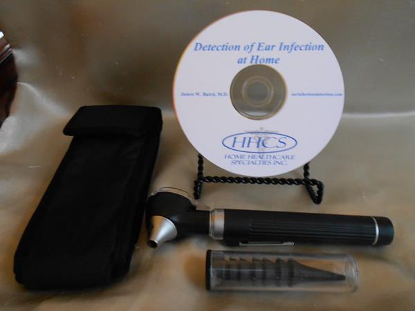
Not to bang my own drum too much and too loudly, you will find a very reasonably priced, lightweight, fiberoptic otoscope with a 3X magnifying viewing window on my website, By accessing this website, you can directly download the digital video file of “Ear Infection Detection at Home”, or purchase the DVD. Also available is a combination product of the DVD and otoscope.
Because I appreciate your business, I am offering a credit in the amount of $5.00 on any product on my site, with the exception of the direct digital download. Utilize the following credit code to claim your credit: EBOOK
Thank you so much for downloading this ebook. Use the information contained herein, or in the digital download/DVD, as intended. That is, use it as a tool to help you make a more informed decision about your child’s or family’s healthcare needs.
James W. Baird, M.D., graduated from the University of Texas Medical Branch in 1974. His early career was spent in Emergency Medicine and Urgent Care. He now practices in a primary care setting.
International Criminal Court, Article 98(2) and Bilateral Immunity Agreement Dinesh Tripathi Postal Address: P.O. Box 19186, Kathmandu, Nepal Tel: +977-1-44 23 125, Fax: +977-1-44 38 812 International Criminal Court, Article 98(2) and Bilateral Immunity Agreement Overview The establishment of International Criminal Court is a landmark development. It is an inno
Josh Newell, Siberia Hotspot Program, Friends of the Earth - Japan Siberia The term stems from the Tartar word sibir or sleeping land. To foreigners, the name usually refers to the vast stretch of Russia from the Ural Mountains in the west to the Pacific seacoast in the east. Russians, however, generally consider Siberia’s eastern edge to be a series of mountain ranges stretching from w
 HOME DETECTION OF EAR INFECTION